
Congenital fibrosis of the extraocular muscles (CFEOM) is a group of rare congenital cranial dysinnervation disorders in which one or more extraocular muscles are replaced by fibrous tissue, producing restrictive, non-progressive ophthalmoplegia present from birth.
It belongs to a broader family of conditions — including Duane retraction syndrome and Möbius syndrome, both discussed elsewhere on this site — now understood to result not from primary muscle disease but from failure of the cranial nerves that should normally innervate these muscles to develop properly, with the muscles themselves undergoing secondary fibrotic change as a consequence of never having received normal innervation.
Reframing the Underlying Mechanism
Although the condition is named for the fibrotic muscle change that is its most visible clinical consequence, the primary defect is now understood to lie in the cranial nerves and their brainstem nuclei — specifically, failure of normal development of the oculomotor and, in some subtypes, trochlear nerve nuclei — rather than in the muscles themselves, which instead fibrose secondarily from a lifetime of absent or abnormal innervation starting in utero.
This reframing, from a primary myopathy to a primary neurogenic disorder, is the modern understanding that unifies CFEOM with the other congenital cranial dysinnervation disorders as a shared category of developmental miswiring rather than a group of unrelated conditions that happen to share superficial clinical similarities.
Classification
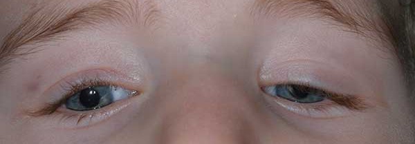
- CFEOM1 — the most common and most severe form, caused by KIF21A mutations, producing bilateral, severe ptosis with the eyes fixed in a markedly downward position and severely restricted movement in essentially all directions
- CFEOM2 — caused by PHOX2A mutations, autosomal recessive, generally producing bilateral ptosis with the eyes fixed in an outward (exotropic) position
- CFEOM3 — genetically and phenotypically more variable (including TUBB3 and other mutations), sometimes unilateral or asymmetric, and generally somewhat milder than CFEOM1
Each subtype has a characteristic, though not perfectly rigid, pattern of eye position and severity.
Some CFEOM3 cases are associated with additional neurologic features beyond the eye findings, reflecting the broader neurodevelopmental role of some of the implicated genes.
All-fit Slit-Lamp Adapter
Record and share exactly what you see at the slit lamp. One adapter fits any slit lamp or surgical microscope — and any smartphone.
From Choroida — the team behind this siteClinical Presentation
- Bilateral (or, in some CFEOM3 cases, unilateral) ptosis, present from birth and typically severe enough to be immediately apparent
- The eyes are fixed in an abnormal position (commonly downward in CFEOM1, outward in CFEOM2), with severely restricted voluntary movement in most or all directions of gaze
- A characteristic compensatory chin-up head posture, adopted from infancy to bring the visual axis out from under the ptotic lids and away from the abnormally fixed eye position, allowing the child to see beneath the drooping eyelids
- Absence of Bell’s phenomenon (the normal reflexive upward eye rotation on attempted forced lid closure) in many patients, given how limited overall eye movement is
- A stable, non-progressive course from birth — this is a genuinely important distinguishing feature, since a truly progressive ophthalmoplegia in later childhood or adulthood should prompt reconsideration of the diagnosis rather than being attributed to CFEOM
Differential Diagnosis
- Congenital ptosis without associated restrictive ophthalmoplegia — isolated levator dysfunction without the fixed, restricted eye position characteristic of CFEOM
- Duane retraction syndrome — a related congenital cranial dysinnervation disorder, but with its own distinct pattern (globe retraction on adduction, horizontal rather than the more complex multi-directional restriction of CFEOM), discussed in relation to other conditions in this same disorder family elsewhere on this site
- Chronic progressive external ophthalmoplegia — a mitochondrial disorder producing progressive, acquired (rather than congenital and static) ophthalmoplegia, discussed in its own dedicated article on this site; the progressive course and typically later onset are the key distinguishing features from CFEOM
- Myasthenia gravis — acquired, variable, fatigable weakness rather than a fixed, congenital restrictive pattern, discussed in this site’s coverage of ocular myasthenia gravis
Diagnostic Evaluation
The diagnosis is largely clinical, based on the characteristic congenital, stable, restrictive ophthalmoplegia pattern with associated ptosis.
Forced duction testing confirms the restrictive (rather than paretic) nature of the limited eye movement, similar in principle to the confirmatory role forced duction testing plays in diagnosing Brown syndrome, discussed in its own dedicated article on this site.
Genetic testing for the relevant CFEOM genes confirms the specific subtype and supports genetic counseling for the family, given the syndrome’s varying inheritance patterns across the different subtypes (predominantly autosomal dominant for CFEOM1 and CFEOM3, autosomal recessive for CFEOM2).
Management
Because the underlying nerve and muscle abnormality cannot be reversed, management focuses on optimizing functional vision and addressing the ptosis and abnormal head posture: ptosis surgery (typically frontalis sling procedures, given how poor native levator function usually is in this condition) to lift the eyelids out of the visual axis, and strabismus surgery to bring the eyes into a more useful position and reduce the need for an extreme compensatory head posture.
Amblyopia surveillance and treatment is essential in affected children, given the combination of significant ptosis and abnormal eye position, both of which carry real amblyogenic risk if not actively monitored and addressed during the critical period of visual development.
Because these children often need staged surgery over several years — ptosis correction, strabismus repair, and occasionally repeat procedures as they grow — ongoing coordinated follow-up with pediatric ophthalmology is essential rather than a single intervention at diagnosis.


Document what you see
Two smartphone imaging tools built for everyday clinic use — one for the slit lamp, one for the fundus.
From Choroida — the team behind this siteReferences
- Engle EC. Genetic basis of congenital strabismus. Archives of Ophthalmology.
- Yamada K, Andrews C, Chan WM, et al. Heterozygous mutations of the kinesin KIF21A in congenital fibrosis of the extraocular muscles type 1 (CFEOM1). Nature Genetics.
- American Academy of Ophthalmology. Basic and Clinical Science Course, Section 6: Pediatric Ophthalmology and Strabismus.
Test yourself
A few questions straight from this article.
-
Where does the primary defect lie in congenital fibrosis of the extraocular muscles (CFEOM)?
Failure of normal oculomotor, and in some subtypes trochlear, nuclear development is the primary defect; the muscles fibrose secondarily from a lifetime of absent innervation. -
CFEOM belongs to which group of disorders?
CFEOM sits alongside Duane retraction syndrome and Mobius syndrome as a disorder of developmental miswiring of the cranial nerves rather than primary muscle disease. -
Which gene is mutated in CFEOM1, the most common and most severe form?
CFEOM1 results from KIF21A mutations and produces bilateral severe ptosis with the eyes fixed markedly downward and movement restricted in essentially all directions. -
Which CFEOM subtype is autosomal recessive with the eyes fixed in an outward position?
CFEOM2 is caused by PHOX2A mutations, is inherited in an autosomal recessive manner, and typically produces bilateral ptosis with exotropic eyes. -
A child diagnosed with CFEOM develops clearly worsening ophthalmoplegia years later. What does this indicate?
CFEOM runs a stable, non-progressive course from birth, so genuinely progressive ophthalmoplegia should prompt reconsideration rather than being attributed to CFEOM. -
Why do children with CFEOM adopt a chin-up head posture from infancy?
The chin-up posture lets the child see beneath drooping eyelids and compensates for the abnormally fixed eye position. -
Which feature distinguishes chronic progressive external ophthalmoplegia from CFEOM?
Chronic progressive external ophthalmoplegia is a mitochondrial disorder that is acquired and progressive, whereas CFEOM is congenital and static. -
What does forced duction testing establish in a child with suspected CFEOM?
Forced duction testing confirms the restrictive nature of the limitation, the same confirmatory role it plays in Brown syndrome. -
Why is a frontalis sling typically chosen for ptosis surgery in CFEOM?
Because levator function in CFEOM is generally poor, frontalis sling procedures are used to lift the lids out of the visual axis. -
Which ongoing task is essential in children with CFEOM during visual development?
Significant ptosis combined with an abnormal eye position carries real amblyogenic risk, so active monitoring and treatment through the critical period is essential.
