
Case Study
A 72-year-old female with a known history of neovascular age-related macular degeneration (nAMD) presented for follow-up.
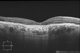
She reported a gradual decline in central vision in her left eye despite ongoing anti-VEGF injections. On spectral-domain optical coherence tomography (SD-OCT), multiple tubular, hyporeflective structures with hyperreflective borders were observed within the outer retina, particularly in areas adjacent to regions of geographic atrophy.
These structures were consistent with outer retinal tubulation (ORT). Fluorescein angiography did not reveal active leakage, and intraocular pressure was within normal limits.
A diagnosis of outer retinal tubulation associated with chronic nAMD was made.
Disease Entity
Outer Retinal Tubulation (ORT) is a structural retinal change observed primarily in chronic degenerative retinal diseases.
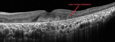
It represents a response to photoreceptor injury, where surviving photoreceptors rearrange themselves into tubular formations within the outer nuclear layer.
ORT is frequently seen in association with age-related macular degeneration (AMD), particularly in advanced stages, and also occurs in retinitis pigmentosa, choroideremia, Stargardt disease, and other inherited retinal dystrophies.
Fundus Explorer Pro
Photograph the retinal findings described here with the phone already in your pocket — 22 D optics and built-in illumination in one handheld unit.
From Choroida — the team behind this sitePathophysiology
ORT is thought to arise from reactive reorganization of degenerating photoreceptors. As the surrounding support structures—especially the retinal pigment epithelium (RPE) and choriocapillaris—degenerate, photoreceptor cells attempt to maintain structural integrity by forming these circular or ovoid tubulations.
Histologically, these structures are composed of degenerating cone and rod photoreceptors, Müller cells, and reorganized outer retinal material.
They are distinct from cystoid spaces in that they do not represent fluid accumulation, but rather, degenerative tissue remodeling.
Epidemiology
-
ORT is increasingly identified due to widespread OCT usage.
-
Common in late-stage AMD, particularly geographic atrophy and disciform scars.
-
Seen in ~20–30% of eyes with neovascular AMD undergoing anti-VEGF treatment.
-
Also identified in inherited retinal degenerations, high myopia, ocular toxoplasmosis, and central serous chorioretinopathy.
Clinical Features
Symptoms:
-
ORT itself is asymptomatic.
-
Vision loss, if present, is typically due to the underlying pathology, not ORT.
Signs:
-
OCT features:
-
Round or ovoid hyporeflective spaces with hyperreflective borders.
-
Located in the outer nuclear layer (ONL) or at the junction of the outer plexiform layer (OPL).
-
Frequently found adjacent to areas of atrophy, fibrosis, or scarring.
-
-
No leakage was seen on fluorescein angiography.
-
Static structures: ORTs do not change or respond to anti-VEGF therapy.
Examination Findings
-
Visual Acuity: Variable; depends on the underlying disease.
-
Fundus Examination: Usually normal or showing signs of underlying retinal pathology.
-
OCT:
-
Hyporeflective tubular structures within the ONL.
-
Hyperreflective outer walls.
-
Located at the junction of atrophic or fibrotic zones.
-
-
Fluorescein Angiography (FA):
-
No leakage.
-
-
Indocyanine Green Angiography (ICGA):
-
May show choroidal thinning or polypoidal lesions in associated diseases.
-
Differential Diagnosis
It is crucial to differentiate ORT from intraretinal cystoid spaces, as their management and implications differ.
| Feature | ORT | Cystoid Spaces |
|---|---|---|
| Reflectivity | Hyporeflective center with hyperreflective wall | Hyporeflective without border |
| Location | Outer retina (ONL) | Inner retina |
| Shape | Ovoid/tubular | Round/oval |
| Leakage on FA | No | Often present |
| Associated with | Chronic degeneration | Active exudation/inflammation |
Diagnosis
ORT is diagnosed based on OCT imaging, with hallmark findings of:
-
Tubular structures with distinct hyperreflective walls.
-
Stable appearance over time.
-
Located in the outer retinal layers.
-
Adjacent to areas of geographic atrophy, fibrosis, or scarring.
No invasive testing is typically required, though fundus autofluorescence and angiography may aid in evaluating concurrent pathologies.
Management
ORT does not require treatment. Instead, the focus is on managing the underlying retinal condition.
Key management principles:
-
Avoid misdiagnosis as cystoid macular edema or exudation to prevent unnecessary treatments.
-
Continue regular monitoring for progression of the primary disease (e.g., AMD).
-
Consider genetic counseling in inherited dystrophies.
-
Use low-vision aids and vision rehabilitation for advanced visual loss.

Prognosis
-
ORT itself is benign and non-progressive.
-
Its presence, however, typically indicates advanced disease and correlates with poorer visual prognosis due to photoreceptor degeneration.
-
Recognition is essential to avoid overtreatment and misinterpretation of imaging studies.
Prevention
-
ORT cannot be prevented directly.
-
Regular monitoring and early intervention in the primary disease may reduce the severity or delay the development of ORT.
-
Optimizing retinal health and managing systemic risk factors (e.g., hypertension, hypercholesterolemia) in AMD may indirectly help.
Conclusion
Outer Retinal Tubulation is a structural retinal remodeling process resulting from chronic photoreceptor degeneration.
While benign in itself, its identification is crucial as a marker of disease chronicity and advanced retinal damage.
With the rise in high-resolution OCT imaging, ORT is now more frequently recognized.
Understanding its features helps ophthalmologists avoid unnecessary treatments, guide patients effectively, and focus care on the underlying condition.


Document what you see
Two smartphone imaging tools built for everyday clinic use — one for the slit lamp, one for the fundus.
From Choroida — the team behind this siteReferences
-
Zweifel SA, Spaide RF, et al. Outer Retinal Tubulation in Advanced Age-Related Macular Degeneration: Optical Coherence Tomographic Findings. Ophthalmology. 2009.
-
Dolz-Marco R, Gal-Or O, Freund KB, et al. Outer retinal tubulation and its relationship to subretinal fibrosis in neovascular age-related macular degeneration. Am J Ophthalmol. 2017.
-
Keane PA, Sadda SR. Predicting optical coherence tomography patterns in retinal disease. Retina. 2011.
-
Zweifel SA, Engelbert M, Laud K, et al. Outer retinal tubulation: a novel optical coherence tomography finding. Arch Ophthalmol. 2009.
-
Litts KM, Zhang Y, Freund KB, et al. Inner segment remodeling and mitochondrial degeneration are early events in outer retinal tubulation. Acta Neuropathol Commun. 2015.
-
Giannakaki-Zimmermann H, Ebneter A, Wolf S, Zinkernagel MS. Outer retinal tubulation in age-related macular degeneration: longitudinal analysis and comparison with other retinal conditions. Br J Ophthalmol. 2016.
Test yourself
A few questions straight from this article.
-
Outer retinal tubulation represents which structural process in chronic retinal disease?
ORT is a reactive response to photoreceptor injury in which surviving photoreceptors reorganise themselves into tubular formations within the outer nuclear layer. -
Which cells contribute histologically to outer retinal tubulation alongside degenerating photoreceptors?
Histologically ORT is composed of degenerating cone and rod photoreceptors, Müller cells, and reorganised outer retinal material — not fluid. -
Roughly what proportion of eyes with neovascular AMD on anti-VEGF therapy show outer retinal tubulation?
ORT is seen in approximately 20-30% of eyes with neovascular AMD undergoing anti-VEGF treatment, and is increasingly recognised with widespread OCT use. -
What is the hallmark OCT appearance of outer retinal tubulation?
ORT appears on OCT as round or ovoid hyporeflective spaces bounded by distinct hyperreflective walls, sited in the outer nuclear layer or at the outer plexiform junction. -
Where in the retina is outer retinal tubulation located on OCT?
ORT sits in the outer nuclear layer, or at the junction with the outer plexiform layer, typically adjacent to zones of atrophy, fibrosis or scarring. -
Which feature best distinguishes outer retinal tubulation from intraretinal cystoid spaces?
ORT shows a hyporeflective centre with a hyperreflective wall and never leaks on angiography, whereas cystoid spaces are hyporeflective without a border and often leak. -
How do outer retinal tubulations behave on serial imaging during anti-VEGF therapy?
ORTs are static structures that do not change or respond to anti-VEGF therapy, which is why mistaking them for exudation leads to unnecessary injections. -
What is the recommended management of outer retinal tubulation itself?
ORT requires no treatment. Care focuses on the underlying condition, with regular monitoring and avoidance of misdiagnosis as cystoid macular oedema or exudation. -
Besides AMD, in which group of disorders is outer retinal tubulation commonly identified?
ORT occurs in retinitis pigmentosa, choroideremia, Stargardt disease and other inherited dystrophies, as well as in high myopia, ocular toxoplasmosis and central serous chorioretinopathy. -
What does the presence of outer retinal tubulation imply about visual prognosis?
ORT is benign and non-progressive in itself, but its presence indicates advanced disease and correlates with a poorer visual prognosis from photoreceptor degeneration.
