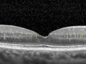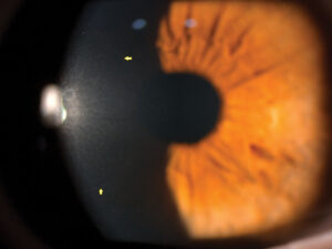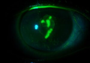
Slit lamp Exam is the most important part to detect any abnormality or disease during eye examination.
How to do a slit lamp exam
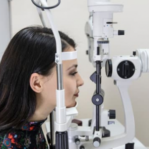
- Patient should be positioned comfortably with his chin resting on chin rest and forehead opposed to head rest.
- The height of the table housing the slit lamp should be adjusted according to patient’s height.
- The microscope and illumination system should be aligned with the patient’s eye to be examined.
- Focus the microscope by
- Adjusting the interpupillary distance
- Adjusting the eyepieces (set at 0 or dial in your refraction)
- Set magnification in on 1x
- Move the microscope up and down, in or out or latterly byjoystickso that the tissue that is of interest comes clearly into view.
- Adjust the illumination or brightness through the desired filters.
- Adjust the width and height of the beam.
Start systematic Slit lamp Exam of eye from front to the back as follow :
1. Eye lid margin
Slit lamp examination beam is widened to a full circle to illuminate the front of eye. Thus the anterior border shows the eyelashes projecting in two or three rows and its posterior border is sharp and resting against theeyeball. There are small orifices of the tarsal glands just in front of the lid margin and the lacrimal papilla.
2. Conjunctiva:
- Palpebral:This structure is seenby everting the eyelids, it is a thin, transparent mucous membrane lining the inner surface of the eyelid.
- Bulbar:This structure is examined by holding the lids open and asking the patient to look to the right, left, up, and down. Here again the conjunctiva is seen as a thin, transparent mucous membrane. A superficial, bright red system of anastomosing vessels can be easily identified in the bulbar conjunctiva.
3. Cornea:
It is the transparent layer. When the slit-lamp beam passes through the cornea, it is possible to recognize the anterior and posterior surfaces and the corneal stroma.
4. Sclera:
slit lamp exam to the sclera is extremely important to check any presence of subconjuctival hge or marks of previous procedures like trabeculotomy for Glaucoma patients
5. Anterior chamber:
Normally the anterior chamber filled with aqueous appears optically empty and black. If the slit beam is narrowed to a fine pencil of light, a very faint relicense can be made out along the course of the light. Angle of anterior chamber is assessed by gonioscopy.
6 .Iris and pupil:
Iris is the colored part of eye. In the centre there is an opening known as pupil. It controls the amount of light going into the eye by the constriction and dilation of pupil.
7. Lens:
Check whether the lens is transparent or cloudy (cataract).
8. Vitreous body
Itis just posterior to the lens. In the vitreous of aged patients, other condensations can be seen; these are thought to represent areas of degeneration.



Document what you see
Two smartphone imaging tools built for everyday clinic use — one for the slit lamp, one for the fundus.
From Choroida — the team behind this siteOther usage of Slit lamp Exam with accessory devices
1. Fundus examination:
Fundus examination can be performed by placing an additional handheld 78D or 90D lens in front of the eye. Normally the fundus has a red appearance, the optic disc is pink, round or oval shaped with sharp margins. Macula is situated 3 mm temporally to the optic disc. It is small circular area, deeper red than the surrounding fundus, there is a bright foveal reflex in the Centre. The normal artery: vein ratio in fundus is 2:3.
2. Applanation tonometry:
The IOP can be measured in slit lamp exam with the help of Goldman applanation tonometer. An applanation tonometer measures the IOP by flattening the cornea overa specific area (3.06mm).
- Method:
Anaesthetize the cornea and stain the tear film with fluorescein. Patient is seated in front of a slit lamp and the cornea and biprisms are illuminated with cobalt blue light from the slit-lamp. Biprism is then advanced until it just touches the apex of cornea. At this point two fluorescent semicircles are viewed through the prisms. The applanation force against the cornea is adjusted until the inner edges of the two semicircles just touch. The IOP is determined by multiplying the dial reading with 10.
3. Gonioscopy:
The angle of the anterior chamber of the eye cannot be seen by direct inspection because of the light rays arising from the angleundergo total internal reflection on reaching the curved surface of the cornea. The use of a special contact lens, a source of focal illumination, and a microscope or slit lamp makes the angle accessible. The contact lens eliminates the corneal curve and allows light to be reflected from the angle.
Slit lamp exam photography
Slit lamp photography can be done by attaching the smartphones to slit-lamp by using slit lamp adaptors. a slit lamp adaptor has two parts, one part holds the smartphone, whereas the other part fixes onto the eyepiece of the slit lamp. There are different types of slit-lamp adaptors according to the eyepiece of the slit-lamp:
- 10x eyepiece adaptors
- 12x eyepiece adaptors

Test yourself
A few questions straight from this article.
-
How should a patient be positioned for a slit lamp exam?
The article specifies comfortable seating with the chin resting on the chin rest and the forehead opposed to the head rest, with table height adjusted to the patient. -
At what magnification does the slit lamp exam article advise starting the microscope?
Focusing the microscope involves setting interpupillary distance, adjusting the eyepieces to 0 or the examiner's refraction, and setting magnification at 1x before moving to the tissue of interest. -
On slit lamp examination, how does the normal anterior chamber appear?
The aqueous-filled anterior chamber is optically empty and black; narrowing the beam to a fine pencil of light shows only a very faint relucence along its course. -
Which lens is held in front of the eye for fundus examination at the slit lamp?
The article states that fundus examination at the slit lamp is performed by placing an additional handheld 78D or 90D lens in front of the eye. -
Where does the slit lamp fundus exam locate the normal macula?
The macula lies about 3 mm temporal to the optic disc as a small circular area deeper red than the surrounding fundus, with a bright foveal reflex at its centre. -
What artery-to-vein ratio is described as normal on fundus examination?
The article gives the normal fundus artery to vein ratio as 2:3, alongside a pink optic disc with sharp margins. -
Over what corneal area does Goldmann applanation tonometry flatten the cornea?
An applanation tonometer derives intraocular pressure from the force needed to flatten a specific corneal area of 3.06 mm. -
How is intraocular pressure obtained from the Goldmann applanation tonometer dial?
Once the endpoint is reached, the article states that intraocular pressure is determined by multiplying the dial reading by 10. -
What is the endpoint when performing applanation tonometry at the slit lamp?
After fluorescein staining and cobalt blue illumination, applanation force is increased until the inner edges of the two fluorescent semicircles just touch. -
Why is the anterior chamber angle invisible on direct slit lamp inspection?
Light rays from the angle are totally internally reflected at the curved corneal surface; a gonioscopy contact lens eliminates that curve and lets the light escape.

Writes and reviews the clinical articles on Educate. Every post is checked against peer-reviewed sources before it goes live.