Retained PFCL is perfluorocarbon liquid that remains in the eye after vitreoretinal surgery.
Surgeons use this heavy liquid to flatten the retina during repair (see perfluorocarbon liquids). However, small bubbles can escape removal and settle under the retina or in the anterior chamber, and although most are harmless, a subfoveal bubble can damage central vision.
What Retained PFCL Is
Perfluoro-n-octane and perfluorodecalin are the agents used most often, and they are clear, dense and immiscible with water. As a result, residual droplets sink to the lowest available point, and their position after surgery therefore depends on head posture and break anatomy.
Where Retained PFCL Settles
Typical locations include:
- Vitreous cavity: shiny droplets resting on the inferior retina
- Subretinal space: a round bubble, often drifting toward the posterior pole
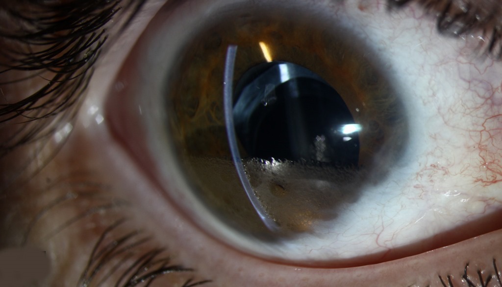
- Anterior chamber: clear spheres layering at the inferior angle
Fundus Explorer Pro
Photograph the retinal findings described here with the phone already in your pocket — 22 D optics and built-in illumination in one handheld unit.
From Choroida — the team behind this siteWhy Bubbles Get Left Behind
Retention usually follows incomplete removal at the end of the case. For example, PFCL can pass through a large break or retinotomy into the subretinal space, and many small droplets, called fish eggs, are also harder to aspirate than one large bubble. Garcia-Valenzuela and colleagues reported that break characteristics and technique influenced the risk.
Symptoms of Retained PFCL
Peripheral droplets usually cause no symptoms, whereas a subfoveal bubble produces a central scotoma or distortion. Floating vitreous droplets may also appear as small round shadows, and anterior chamber droplets rarely bother patients unless the cornea swells.
Fundus Signs of Retained PFCL
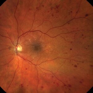
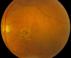
On ophthalmoscopy, the bubbles look like glistening, perfectly round spheres. Subretinal bubbles form sharply outlined, smooth elevations with a bright edge reflex, so document them with color photographs for later comparison.
OCT Appearance of Retained PFCL
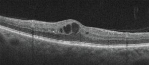
OCT gives the clearest picture, in which a subretinal bubble lifts the neurosensory retina into a smooth, steep dome, and the space beneath looks optically empty and has sharp, regular borders. In contrast, subretinal fluid usually has gentler, sloping margins (see subretinal fluid on OCT). Over time, the outer retina above the bubble can thin, and the ellipsoid zone may fade.
Anterior Chamber PFCL
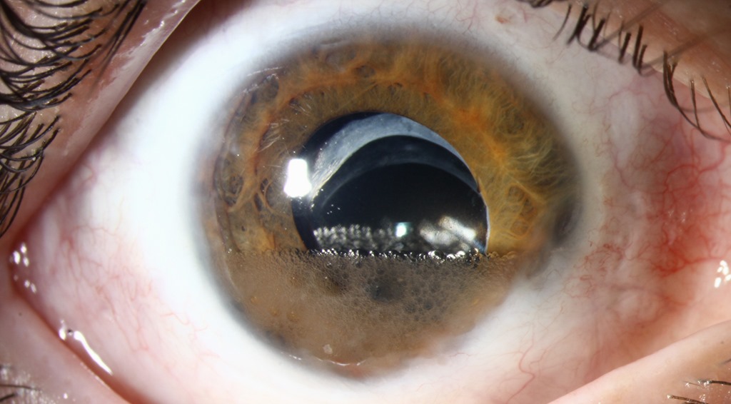
Droplets reach the anterior chamber through a capsular or zonular defect, or in aphakic eyes, and they sit inferiorly, so they can mimic a small hypopyon at a glance. Anterior segment OCT confirms their round, clear profile (see anterior segment OCT), and prolonged contact with the inferior endothelium can also cause localized corneal edema.
Toxicity Concerns With Retained PFCL
The agents are chemically inert in the short term, but prolonged retinal contact appears harmful. Elsing and colleagues described histologic changes, including a cellular reaction, in eyes with retained perfluoro-n-octane, and microperimetry studies also show reduced sensitivity over subretinal bubbles, possibly from mechanical compression as well.
When to Remove Retained PFCL
Removal is generally advised for subfoveal bubbles, because foveal damage may become permanent, and many surgeons also remove bubbles that migrate toward fixation. In the anterior chamber, corneal edema or a large volume favors washout, while small, stable peripheral droplets can usually be observed.
Surgical Options for Subretinal PFCL
Most surgeons aspirate the bubble through a small retinotomy with a fine cannula, and some first inject balanced salt solution under the retina to mobilize it. Roth and colleagues described one such technique for subfoveal perfluoro-n-octane, while gas tamponade with positioning can alternatively displace the bubble away from the fovea. Each approach carries a risk of macular hole, so discuss it openly.
Removing Anterior Chamber PFCL
A paracentesis with gentle irrigation usually clears the droplets. Next, tilt the head so they collect at the wound, and afterward check pressure and endothelial clarity at the next visit.
Timing and Follow-Up of Retained PFCL
Earlier removal seems sensible before chronic outer retinal changes develop, although evidence comes mainly from case series. Repeat OCT shows whether an observed bubble enlarges, migrates or stays stable, and also watch for recurrent detachment, which may change the plan.
Prevention During Surgery
Prevention starts in the operating room, where you first keep the cannula tip inside the bubble so injection builds one large sphere. Then avoid filling PFCL above the level of a peripheral break, and finally aspirate again over the optic disc after a short pause.
Counseling Patients About Retained PFCL
Explain that a small amount of a surgical tool remained in the eye, and reassure patients with peripheral droplets that observation is reasonable. For a subfoveal bubble, weigh the benefit of removal against the risk of macular hole, and also set realistic expectations, because some sensitivity loss may persist.
Pitfalls to Avoid
- Do not mistake anterior chamber PFCL for a hypopyon
- Do not confuse heavy liquid with silicone oil, which floats
- Check the operative notes when a round subfoveal elevation appears
- Request OCT for any new central scotoma after a PFCL case


Document what you see
Two smartphone imaging tools built for everyday clinic use — one for the slit lamp, one for the fundus.
From Choroida — the team behind this siteReferences
- Chang S. Low viscosity liquid fluorochemicals in vitreous surgery. Am J Ophthalmol. 1987;103:38-43.
- Elsing SH, Fekrat S, Green WR, et al. Clinicopathologic findings in eyes with retained perfluoro-n-octane liquid. Ophthalmology. 2001;108:45-48.
- Garcia-Valenzuela E, Ito Y, Abrams GW. Risk factors for retention of subretinal perfluorocarbon liquid in vitreoretinal surgery. Retina. 2004;24:746-752.
- Roth DB, Sears JE, Lewis H. Removal of retained subfoveal perfluoro-n-octane liquid. Am J Ophthalmol. 2004;138:287-289.
- Tewari A, Eliott D, Singh CN, et al. Changes in retinal sensitivity from retained subretinal perfluorocarbon liquid. Retina. 2009;29:248-250.
Test yourself
A few questions straight from this article.
-
Why do retained perfluorocarbon liquid (PFCL) droplets settle at the lowest available point in the eye?
Perfluoro-n-octane and perfluorodecalin are clear, dense and immiscible with water, so residual droplets sink to the lowest point, depending on posture and break anatomy. -
How does PFCL usually reach the subretinal space during vitreoretinal surgery?
Retention usually follows incomplete removal, and PFCL can pass through a large break or retinotomy into the subretinal space. -
During vitrectomy, why do PFCL 'fish eggs' raise the risk of retained heavy liquid?
Many small droplets, called fish eggs, are harder to aspirate than one large bubble, which is why surgeons try to build one large sphere. -
Which symptom suggests a subfoveal retained PFCL bubble after retinal surgery?
Peripheral droplets usually cause no symptoms, whereas a subfoveal bubble produces a central scotoma or distortion. -
On OCT, how does a subretinal PFCL bubble differ from ordinary subretinal fluid?
A subretinal PFCL bubble lifts the retina into a smooth, steep dome with an optically empty space and sharp borders, while subretinal fluid has gentler, sloping margins. -
Retained PFCL layering in the inferior anterior chamber can be mistaken for which finding?
Anterior chamber droplets sit inferiorly and can mimic a small hypopyon; anterior segment OCT confirms their round, clear profile. -
Why is removal generally advised for a subfoveal retained PFCL bubble?
Removal is advised for subfoveal bubbles because foveal damage may become permanent, while small stable peripheral droplets can usually be observed. -
Which complication is a shared risk of the surgical approaches for removing subfoveal PFCL?
Aspiration through a small retinotomy, BSS mobilization or gas displacement each carry a risk of macular hole, so it should be discussed openly. -
Which intraocular agent floats, unlike retained heavy liquid, and should not be confused with it?
Heavy liquid sinks, whereas silicone oil floats, so the two should not be confused when a bubble is seen after surgery. -
What do microperimetry studies show over retained subretinal PFCL bubbles?
Prolonged retinal contact appears harmful: microperimetry shows reduced sensitivity over subretinal bubbles, possibly also from mechanical compression.

Writes and reviews the clinical articles on Educate. Every post is checked against peer-reviewed sources before it goes live.