RPE function covers the daily work of the pigmented cell layer between the photoreceptors and Bruch membrane.
These cells feed, clean and protect the outer retina, so most outer retinal diseases involve the RPE at some stage. The fundus often shows which job has failed.
Anatomy Behind RPE Function
The RPE is a hexagonal monolayer whose apical microvilli wrap the outer segments, and its basal surface rests on Bruch membrane and faces the choriocapillaris. Tight junctions also seal neighboring cells into one sheet, so each cell acts as a polarized gate between two circulations.
Phagocytosis of Outer Segments
Photoreceptors shed the tips of their outer segments every day, and each RPE cell then ingests and digests discs from many photoreceptors. Rod shedding peaks after light onset while cone timing appears more variable, and MERTK, a receptor tyrosine kinase, drives this uptake.
Fundus Explorer Pro
Photograph the retinal findings described here with the phone already in your pocket — 22 D optics and built-in illumination in one handheld unit.
From Choroida — the team behind this siteWhen Phagocytosis Fails
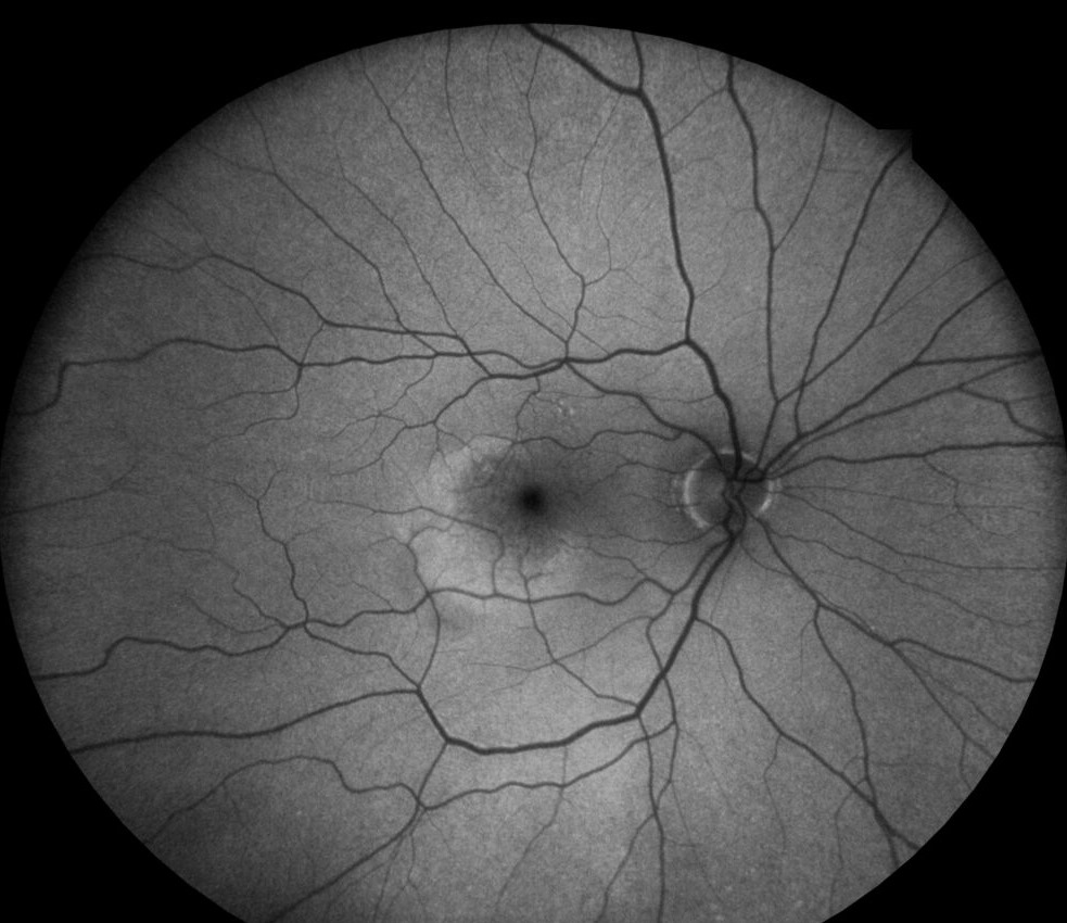
MERTK mutations cause a form of retinitis pigmentosa in humans, and debris then accumulates in the subretinal space while photoreceptors degenerate. Incomplete digestion also leaves lipofuscin, including bisretinoids such as A2E, and autofluorescence maps this load directly (see fundus autofluorescence).
RPE Function in the Visual Cycle
Rods depend on the RPE to regenerate 11-cis retinal after light exposure, and first the RPE takes up all-trans retinol from bleached outer segments. Then LRAT, RPE65 and RDH5 convert it back to 11-cis retinal, and finally CRALBP binds the chromophore before it returns to the photoreceptors.
Visual Cycle Diseases at the Fundus
Each enzyme defect produces a recognizable phenotype.
- RPE65: Leber congenital amaurosis or early-onset severe retinal dystrophy, often with a near-normal early fundus
- RDH5: white dots and slow dark adaptation (see fundus albipunctatus)
- RLBP1: retinitis punctata albescens or Bothnia dystrophy
- Shared symptom: night blindness, because rods rely most on this pathway
Gene Therapy and RPE Function
Voretigene neparvovec delivers a working RPE65 gene by subretinal injection, and in its phase 3 trial, treated patients improved on a multi-luminance mobility test. However, it needs enough viable RPE cells, so advanced atrophy limits benefit. Tell families it may improve dim-light navigation, while normal vision remains out of reach.
The Outer Blood-Retinal Barrier
Tight junctions between RPE cells form the outer blood-retinal barrier and keep leakage from the fenestrated choriocapillaris out of the neural retina. When this seal breaks, fluorescein pools in the subretinal space (see blood-retinal barrier).
RPE Function as a Fluid Pump
The RPE moves water from the subretinal space toward the choroid, and unusually, its Na/K ATPase sits on the apical membrane. Ion transport then draws water across the cell, and this outward flow also helps hold the retina against the RPE.
Barrier and Pump Failure
In central serous chorioretinopathy, a focal RPE leak meets a hyperpermeable choroid, and fluid then collects under the macula as a serous detachment. Similarly, inflammation, tumors and malignant hypertension can overwhelm the pump, and the result is an exudative detachment with shifting fluid.
Melanin and Light Protection in RPE Function
Melanin granules absorb stray light and reduce scatter, and they also bind free radicals and some drugs. In albinism, reduced pigment leaves the choroidal vessels clearly visible, and similarly, focal pigment loss produces a window defect on angiography.
Secretion and the Choriocapillaris
The RPE secretes VEGF from its basal side toward the choriocapillaris, and this signal helps maintain the fenestrated capillaries. Apically, it also releases PEDF, a neuroprotective and antiangiogenic factor, and as a result, choriocapillaris loss tends to follow RPE atrophy.
Ion Buffering and the EOG
The RPE buffers potassium shifts in the subretinal space during light and dark, and its basolateral chloride conductance also generates the light rise of the electrooculogram. In Best vitelliform macular dystrophy, BEST1 mutations reduce this rise, so the Arden ratio falls even when the fundus looks mild.
RPE Function and Macular Degeneration
Age-related macular degeneration shows several RPE jobs failing together, as drusen accumulate between the RPE basement membrane and inner Bruch membrane. Then pigment clumping and RPE loss mark higher progression risk, while in neovascular disease, VEGF imbalance also drives choroidal new vessels.
Reading RPE Function on Imaging
OCT shows the RPE as the outer hyperreflective band, and thinning, elevation or loss of this band localizes the problem. Absent RPE appears dark on autofluorescence, while stressed cells often glow brighter, and fluorescein then separates window defects, blocking and leakage.
Patient Counseling on RPE Function
Many patients hear “pigment layer” and assume a cosmetic problem, so explain that these cells keep the light-sensing cells alive. Then link their symptoms, such as night blindness or distortion, to the failed job, and also offer genetic testing when the pattern suggests an inherited dystrophy.
Pitfalls to Avoid
- Do not dismiss a normal fundus in a child with night blindness
- Do not assume all subretinal fluid comes from neovascularization
- Check the EOG before labeling a vitelliform lesion as AMD
- Remember that dark autofluorescence usually means lost RPE


Document what you see
Two smartphone imaging tools built for everyday clinic use — one for the slit lamp, one for the fundus.
From Choroida — the team behind this siteReferences
- Strauss O. The retinal pigment epithelium in visual function. Physiol Rev. 2005;85:845-881.
- Sparrow JR, Hicks D, Hamel CP. The retinal pigment epithelium in health and disease. Curr Mol Med. 2010;10:802-823.
- Kevany BM, Palczewski K. Phagocytosis of retinal rod and cone photoreceptors. Physiology (Bethesda). 2010;25:8-15.
- Gal A, Li Y, Thompson DA, et al. Mutations in MERTK, the human orthologue of the RCS rat retinal dystrophy gene, cause retinitis pigmentosa. Nat Genet. 2000;26:270-271.
- Russell S, Bennett J, Wellman JA, et al. Efficacy and safety of voretigene neparvovec (AAV2-hRPE65v2) in patients with RPE65-mediated inherited retinal dystrophy. Lancet. 2017;390:849-860.

Writes and reviews the clinical articles on Educate. Every post is checked against peer-reviewed sources before it goes live.