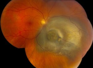
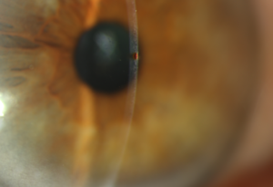
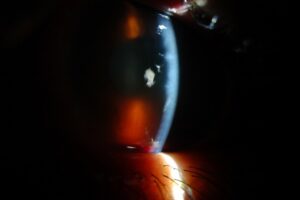
The blood-retinal barrier controls the movement of fluid, ions, and proteins between the blood and the neural retina.
It has an inner and an outer component, and each has its own cells and diseases.
Therefore, understanding it explains why macular edema forms and why fluorescein leaks.
The Inner Blood-Retinal Barrier
Tight junctions between retinal capillary endothelial cells form the inner barrier.
Pericytes, astrocytes, and Müller cell endfeet support the endothelium as a neurovascular unit.
Also, junction proteins such as claudin-5, occludin, and ZO-1 seal the gaps.
Cunha-Vaz and colleagues described this structure and its function in detail.
The Outer Blood-Retinal Barrier
The retinal pigment epithelium forms the outer barrier through tight junctions between its cells.
It separates the leaky choriocapillaris from the outer retina.
Also, the epithelium pumps water and ions toward the choroid and keeps the subretinal space dry (see central serous chorioretinopathy).
Strauss reviewed these transport functions and their importance for vision.
Fundus Explorer Pro
Photograph the retinal findings described here with the phone already in your pocket — 22 D optics and built-in illumination in one handheld unit.
From Choroida — the team behind this siteHow the Blood-Retinal Barrier Breaks Down
VEGF raises permeability by changing the phosphorylation of junction proteins.
Also, hyperglycemia and inflammation damage pericytes and endothelial cells.
Klaassen and colleagues reviewed the molecular basis of this breakdown in diabetes.
As a result, fluid and protein leak into the retina and cause edema.
Diseases of Blood-Retinal Barrier Breakdown
- Diabetic macular edema (see diabetic macular edema)
- Retinal vein occlusion (see central retinal vein occlusion)
- Uveitic macular edema (see uveitic macular edema)
- Postsurgical cystoid macular edema
Clinical Tests of the Blood-Retinal Barrier
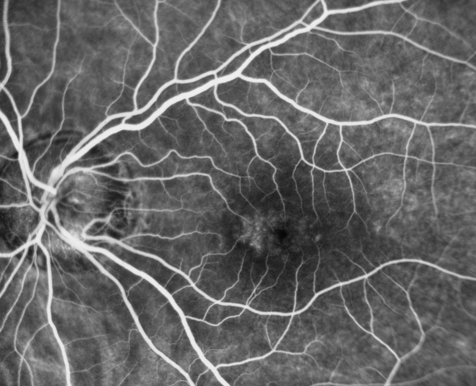
Fluorescein angiography tests barrier integrity, because an intact barrier keeps the dye in the vessels.
Leakage from capillaries signals inner barrier failure, whereas leakage through the epithelium signals outer failure (see fluorescein angiography patterns).
Also, vitreous fluorophotometry measures barrier function quantitatively in research.
Treatment Targets at the Blood-Retinal Barrier
Anti-VEGF drugs reduce permeability by blocking the signal that loosens the junctions.
Corticosteroids restore tight junction proteins and reduce inflammation.
Also, laser may improve the pumping function of the epithelium in selected eyes.
Therefore, each therapy acts on a different part of the barrier.
Why the Barrier Matters for Drug Delivery
The same barrier limits how much drug reaches the retina from the blood.
Therefore, ophthalmologists inject drugs into the eye to bypass it.
Also, new routes such as suprachoroidal delivery aim to improve distribution.
The Neurovascular Unit
The neurovascular unit links neurons, glia, and vessels in one functional group.
Müller cells and astrocytes release factors that tighten the endothelium.
Also, pericytes control capillary tone and support junction stability.
Therefore, damage to any member of the unit can open the barrier.
Why the Choroid Leaks
The choriocapillaris is fenestrated, so plasma proteins cross freely into the choroidal stroma.
Also, the retinal pigment epithelium holds the line against these proteins.
As a result, outer retinal edema appears when the epithelium fails.
This explains the focal leaks in central serous disease.
Aging and Macular Degeneration
With age, Bruch’s membrane thickens and the epithelium loses function.
Also, drusen deposits disturb the transport of nutrients and waste.
Because the outer barrier supports photoreceptors, the damage affects vision.
Therefore, new therapies aim to protect the epithelium.
Role of Inflammation
Cytokines such as TNF-alpha and IL-6 loosen tight junctions.
Also, leukocytes adhere to the endothelium and cause local damage.
As a result, inflammatory eye disease produces macular edema even when vessels are normal.
Corticosteroids work by calming these signals.
Research Tools
Animal models and cell cultures let researchers study barrier function.
Also, imaging with OCT shows the effect of barrier failure as fluid.
Because new methods measure permeability in patients, clinical trials can follow the barrier directly.
Key Points for the Clinician
The barrier keeps the retina dry and the environment stable.
Also, breakdown causes edema in many common diseases.
Because treatments target different parts, match the drug to the mechanism.
Why This Matters for Imaging
Fluorescein angiography and OCT each show the results of barrier failure.
Also, leakage patterns differ between diseases, which helps in diagnosis.
Therefore, learn the anatomy first, and the images make sense.
Barrier Function in Diabetes
Early diabetes damages pericytes and thickens the basement membrane.
Also, the endothelium loses junction proteins, so permeability rises.
As a result, the barrier fails before the vessels show any visible change.
Take-Home Message
Two barriers protect the retina, one at the vessels and one at the pigment epithelium.
Also, each fails in different diseases, so the pattern on imaging tells you where the problem lies.
Therefore, think about the barrier whenever you meet edema.


Document what you see
Two smartphone imaging tools built for everyday clinic use — one for the slit lamp, one for the fundus.
From Choroida — the team behind this siteReferences
- Cunha-Vaz JG. The blood-ocular barriers. Surv Ophthalmol. 1979;23:279-296.
- Cunha-Vaz J, Bernardes R, Lobo C. Blood-retinal barrier. Eur J Ophthalmol. 2011;21(Suppl 6):S3-S9.
- Klaassen I, Van Noorden CJ, Schlingemann RO. Molecular basis of the inner blood-retinal barrier and its breakdown in diabetic macular edema and other pathological conditions. Prog Retin Eye Res. 2013;34:19-48.
- Strauss O. The retinal pigment epithelium in visual function. Physiol Rev. 2005;85:845-881.

Writes and reviews the clinical articles on Educate. Every post is checked against peer-reviewed sources before it goes live.