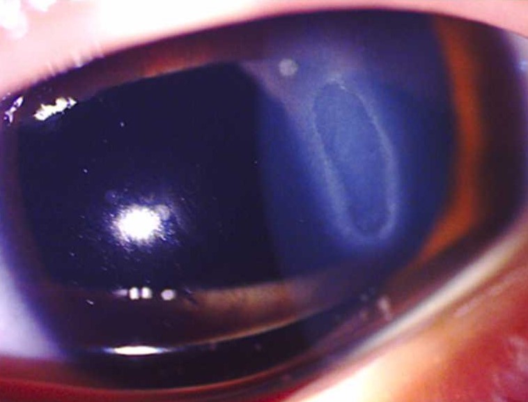
Shield ulcer is a sterile, oval epithelial defect of the superior cornea in vernal keratoconjunctivitis (VKC).
It usually affects boys with active tarsal disease and giant papillae. Because it threatens vision in children, every case needs prompt recognition and a clear plan.
What a Shield Ulcer Looks Like
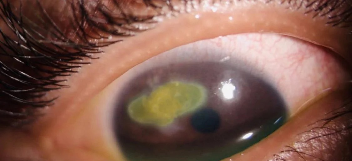
The lesion sits in the upper third of the cornea, often above the visual axis. It is a shallow, horizontally oval defect with well-defined edges. Fluorescein stains the base, while the nearby cornea often shows punctate epitheliopathy, and the patient often has thick mucus and active upper tarsal papillae.
Why the Shield Ulcer Forms
Two mechanisms act together: first, eosinophils release major basic protein and other toxic mediators onto the epithelium, and then giant papillae rub the superior cornea with every blink. As a result, punctate erosions coalesce into a macroerosion and finally a frank ulcer.
All-fit Slit-Lamp Adapter
Record and share exactly what you see at the slit lamp. One adapter fits any slit lamp or surgical microscope — and any smartphone.
From Choroida — the team behind this sitePlaque Formation in Shield Ulcer
Fibrin, mucus and inflammatory debris can collect on the ulcer base and form a gray or white plaque that rises above the corneal surface. The plaque blocks epithelial migration from the edges, so healing stalls even when inflammation improves.
Symptoms Patients Report
Children describe pain, photophobia, tearing and a persistent foreign body sensation, and many keep their eyes closed and miss school. Vision drops when the ulcer or its scar reaches the pupil, although itching usually dominates the history before the ulcer appears.
Slit Lamp Examination of a Shield Ulcer
Evert the upper lid in every child with VKC and a corneal complaint, then grade the papillae and look for Trantas dots at the limbus. Next, study the ulcer base with a narrow slit to judge clarity, elevation and depth, and finally check for stromal infiltrate, anterior chamber reaction and thinning.
Cameron Grading of Shield Ulcer
Cameron proposed a three-grade system in 1995 based on the appearance of the ulcer base.
- Grade 1: transparent base, with good re-epithelialization and a favorable outcome
- In grade 2, a translucent base with deposits delays healing and raises the risk of complications
- Grade 3: an elevated plaque that responds poorly to medical therapy and usually needs surgery
Therefore, the grade tells you early whether drops alone are likely to work.
Differential Diagnosis
- Infectious keratitis, especially with infiltrate or hypopyon
- Herpes simplex epithelial keratitis after steroid exposure
- Atopic keratoconjunctivitis in older patients (see atopic keratoconjunctivitis and the shield ulcer)
- Traumatic abrasion or exposure keratopathy
A superior location with active papillae and a sterile base favors the diagnosis.
Medical Treatment of Shield Ulcer
First, control the allergic drive with topical corticosteroids at a frequent dose, while cyclosporine or tacrolimus helps maintain control and reduces steroid exposure. In addition, a topical antibiotic protects the open epithelium from infection. Mast cell stabilizers and antihistamines support therapy, although they cannot heal an established ulcer alone.
Steroid Risks in Children
Prolonged steroid use raises intraocular pressure and can cause cataract, and it also increases the risk of bacterial and herpetic infection. Therefore, check pressure at every visit and taper once the epithelium closes.
Plaque Debridement in Shield Ulcer
A plaque acts as a physical barrier to epithelial healing, so remove it when it persists despite treatment or when the ulcer reaches grade 3. The surgeon lifts the plaque with a blade or forceps under the microscope, often as a superficial keratectomy, and then the epithelium can slide across a smooth base.
Amniotic Membrane for Shield Ulcer
Amniotic membrane transplantation is an option for ulcers that fail to heal after debridement, and it supports epithelial migration and may dampen surface inflammation. Case series suggest benefit in higher grades, although controlled data remain limited.
Treating the Source: Giant Papillae
The upper tarsus keeps damaging the cornea until it calms down, and supratarsal corticosteroid injection can shrink large papillae in resistant cases. Surgeons excise papillae only in refractory disease, but papillae can recur, so medical control must continue.
Complications of Shield Ulcer
- Secondary bacterial keratitis (see ulcer vs abrasion)
- Subepithelial scar in the superior cornea
- Irregular astigmatism
- Amblyopia in young children
- Steroid glaucoma and cataract
Counseling Families About Shield Ulcer
Explain that the ulcer is sterile but still needs close review every few days. Parents should stop eye rubbing and keep every follow-up visit, and you should also warn them that VKC flares in warm seasons and that ulcers can return.
Most children improve as VKC becomes less active after puberty, while scars may remain. After healing, refract the child and treat amblyopia when the scar affects vision.
Pitfalls to Avoid
- Do not skip lid eversion in a child with a superior corneal defect
- Never rely on medical therapy alone for a persistent plaque
- Avoid stopping steroids abruptly once the ulcer closes
- Check for infection under a sterile-looking plaque


Document what you see
Two smartphone imaging tools built for everyday clinic use — one for the slit lamp, one for the fundus.
From Choroida — the team behind this siteReferences
- Cameron JA. Shield ulcers and plaques of the cornea in vernal keratoconjunctivitis. Ophthalmology. 1995;102:985-993.
- Reddy JC, Basu S, Saboo US, et al. Management, clinical outcomes, and complications of shield ulcers in vernal keratoconjunctivitis. Am J Ophthalmol. 2013;155:550-559.
- Bonini S, Bonini S, Lambiase A, et al. Vernal keratoconjunctivitis revisited: a case series of 195 patients with long-term followup. Ophthalmology. 2000;107:1157-1163.
- Trocme SD, Kephart GM, Bourne WM, et al. Eosinophil granule major basic protein deposition in corneal ulcers associated with vernal keratoconjunctivitis. Am J Ophthalmol. 1993;115:640-643.
- Bowling B. Kanski’s Clinical Ophthalmology: A Systematic Approach. 8th ed. Elsevier; 2016.

Writes and reviews the clinical articles on Educate. Every post is checked against peer-reviewed sources before it goes live.