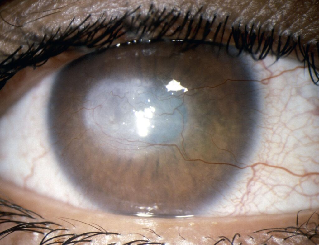
Corneal scar treatment depends on two findings: how deep the opacity lies and whether it crosses the visual axis.
However, a dense central scar can reduce vision through opacity, irregular astigmatism, or both. Therefore, management follows a ladder from optical correction to full-thickness grafting.
Why Scars Blur Vision
A corneal scar degrades vision by two separate mechanisms. First, stromal opacity scatters light and lowers contrast, and second, scar contraction and thinning distort the anterior surface. As a result, a faint scar with marked irregularity may blur more than a dense peripheral one.
Assessment Before Corneal Scar Treatment
Start with slit lamp biomicroscopy in a narrow beam to judge scar depth, then use anterior segment OCT to measure the depth of the opacity and the residual stromal thickness. Topography or tomography also shows how much irregular astigmatism the scar induces. In addition, record endothelial cell density with specular microscopy when the posterior cornea looks healthy.
All-fit Slit-Lamp Adapter
Record and share exactly what you see at the slit lamp. One adapter fits any slit lamp or surgical microscope — and any smartphone.
From Choroida — the team behind this siteEstimating Potential Vision
Before any surgery, ask how much vision the eye can actually recover, for which a rigid lens over-refraction is the most practical test because it neutralizes surface irregularity. If acuity improves sharply, irregular astigmatism drives the loss.
Then check the retina and optic nerve, using B-scan ultrasound when the scar hides the fundus. In children, also look for amblyopia, since a clear cornea will not reverse an established deficit.
Timing of Corneal Scar Treatment
Scars remodel for months after the injury or infection settles, and therefore most surgeons wait until the eye has been quiet for several months before planning keratoplasty. Active inflammation, epithelial defects and infection should resolve first.
Rigid and Scleral Lenses in Corneal Scar Treatment
Rigid gas permeable lenses create a smooth optical surface over an irregular cornea, while scleral lenses vault the whole cornea and suit eyes that cannot hold a corneal lens. For mild opacity with marked irregularity, this step often restores useful vision without surgery. However, lenses cannot clear a dense opacity, and some patients cannot handle them.
PTK in Corneal Scar Treatment
Phototherapeutic keratectomy uses the excimer laser to ablate superficial opacities, and it suits scars confined to the anterior stroma, with enough thickness left after ablation. Masking fluid fills surface valleys, so the laser smooths peaks before it deepens the floor, and in addition, many surgeons apply mitomycin C to reduce postoperative haze.
Limits and Risks of PTK
Removing central tissue flattens the cornea and often induces a hyperopic shift, and haze can recur, especially after deep ablation. Herpes simplex keratitis may reactivate after laser treatment, so oral antiviral cover is sensible in these eyes. Also warn patients that PTK cannot treat deep stromal or full-thickness scars.
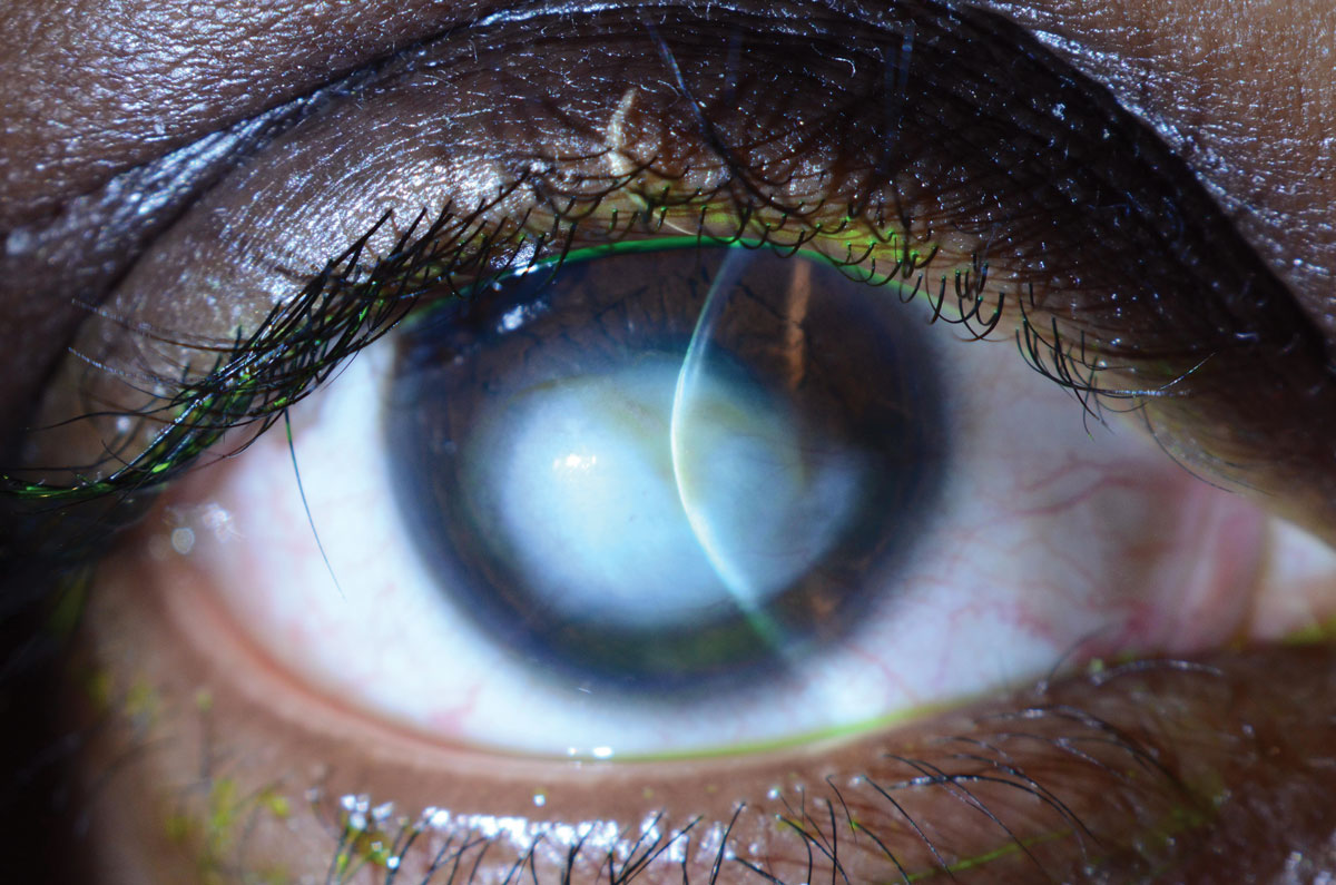
Lamellar Grafts in Corneal Scar Treatment
When the opacity extends into mid or deep stroma, lamellar keratoplasty becomes the next step, and anterior lamellar grafts replace diseased stroma and keep the host endothelium. Automated or manual anterior lamellar keratoplasty suits scars in the anterior half of the stroma, while deeper scars need deep anterior lamellar keratoplasty (see DALK and its indications).
DALK Technique and Evidence
The big-bubble technique injects air to separate stroma from Descemet membrane, and as a result, the surgeon removes nearly all scarred stroma while the host endothelium stays. An American Academy of Ophthalmology review found visual outcomes comparable to penetrating keratoplasty when dissection is deep.
Limits of DALK
Scars that reach Descemet membrane make the dissection harder, and microperforation is common in deep traumatic or post-infectious scars and may force conversion. Therefore, consent patients for possible full-thickness grafting before surgery.
Penetrating Keratoplasty
Full-thickness scars, prior perforations and endothelial failure all point to penetrating keratoplasty, which replaces every corneal layer and clears the optical path in one step (see penetrating keratoplasty). However, it carries lifelong risks of endothelial rejection, wound dehiscence and high astigmatism.
High-Risk Eyes and Corneal Scar Treatment
Vascularized, previously grafted or herpetic eyes carry a higher rejection risk, and herpetic scars also need long-term oral antiviral prophylaxis after keratoplasty. Chemical injury with limbal stem cell deficiency needs surface restoration before a graft can survive.
Counseling on Corneal Scar Treatment
Explain which mechanism limits vision, because this predicts the benefit of each step, and patients should know that grafts need years of follow-up and steroid drops. Then classify the scar clearly to set expectations (see nebula, macula and leucoma).
Pitfalls to Avoid
- Do not graft a cornea before testing a rigid lens over-refraction
- Never plan PTK without measuring scar depth on OCT
- Do not ignore amblyopia in children with dense scars
- Avoid laser or surgery in herpetic eyes without antiviral cover
- Do not rush keratoplasty while the eye is still inflamed


Document what you see
Two smartphone imaging tools built for everyday clinic use — one for the slit lamp, one for the fundus.
From Choroida — the team behind this siteReferences
- Fagerholm P. Phototherapeutic keratectomy: 12 years of experience. Acta Ophthalmol Scand. 2003;81:19-32.
- Anwar M, Teichmann KD. Big-bubble technique to bare Descemet’s membrane in deep anterior lamellar keratoplasty. J Cataract Refract Surg. 2002;28:398-403.
- Reinhart WJ, Musch DC, Jacobs DS, et al. Deep anterior lamellar keratoplasty as an alternative to penetrating keratoplasty: a report by the American Academy of Ophthalmology. Ophthalmology. 2011;118:209-218.
- Tan DT, Dart JK, Holland EJ, Kinoshita S. Corneal transplantation. Lancet. 2012;379:1749-1761.
- American Academy of Ophthalmology. Basic and Clinical Science Course, Section 8: External Disease and Cornea. San Francisco: AAO; 2023.
Test yourself
A few questions straight from this article.
-
Which two findings determine the choice of corneal scar treatment?
Management depends on how deep the opacity lies and whether it crosses the visual axis, and then follows a ladder from optical correction to full-thickness grafting. -
By which two mechanisms does a corneal scar degrade vision?
Stromal opacity scatters light and lowers contrast, while scar contraction and thinning distort the surface, so a faint but irregular scar may blur more than a dense peripheral one. -
What is the most practical test of potential vision before corneal scar surgery?
A rigid lens over-refraction neutralizes surface irregularity; if acuity improves sharply, irregular astigmatism rather than opacity drives the loss, so no cornea should be grafted before this test. -
Which test measures corneal scar depth and residual stromal thickness before treatment?
After narrow-beam slit lamp assessment, anterior segment OCT measures opacity depth and residual stromal thickness; PTK should never be planned without it. -
In a child with a dense corneal scar, which condition must be sought alongside planning surgery?
Children should be checked for amblyopia, because a clear cornea will not reverse an established deficit. -
Which corneal scars are suitable for phototherapeutic keratectomy?
PTK uses the excimer laser to ablate superficial opacities and suits scars in the anterior stroma with enough thickness left after ablation; it cannot treat deep or full-thickness scars. -
Which refractive change commonly follows phototherapeutic keratectomy for a central corneal scar?
Removing central tissue flattens the cornea and often induces a hyperopic shift; haze can also recur, especially after deep ablation. -
What precaution is needed before laser or keratoplasty for a herpetic corneal scar?
Herpes simplex keratitis may reactivate after laser treatment, and herpetic scars need long-term oral antiviral prophylaxis after keratoplasty. -
In the big-bubble DALK technique for corneal scars, what does the injected air achieve?
Air separates stroma from Descemet membrane so nearly all scarred stroma is removed while the host endothelium stays; outcomes approach penetrating keratoplasty when dissection is deep. -
Which corneal scar finding points to penetrating keratoplasty rather than lamellar grafting?
Full-thickness scars, prior perforations and endothelial failure point to penetrating keratoplasty, which replaces every layer but carries lifelong rejection, dehiscence and astigmatism risks.

Writes and reviews the clinical articles on Educate. Every post is checked against peer-reviewed sources before it goes live.