Retinal blood supply comes from two separate circulations that meet near the outer plexiform layer.
The central retinal artery feeds the inner retina, while the choroid nourishes the photoreceptors. Because each system fails in its own way, the fundus picture often identifies the occluded vessel.
Two Systems in Retinal Blood Supply
The retina has one of the highest oxygen demands of any tissue in the body, so it relies on two vascular beds with different structure and control. The retinal circulation serves roughly the inner two thirds of the retina, while the choroid supplies the outer retina, including the photoreceptors and retinal pigment epithelium.
Central Retinal Artery Territory
The central retinal artery enters the optic nerve about one centimeter behind the globe, then emerges on the disc and divides into superior and inferior branches. Next, these divide into nasal and temporal arterioles that respect the horizontal raphe. For example, an embolus in the superotemporal arteriole whitens only that quadrant (see branch retinal artery occlusion).
Fundus Explorer Pro
Photograph the retinal findings described here with the phone already in your pocket — 22 D optics and built-in illumination in one handheld unit.
From Choroida — the team behind this siteCapillary Plexuses of the Inner Retina
Retinal arterioles feed layered capillary networks in the macula, and the superficial plexus lies in the nerve fiber and ganglion cell layers. Intermediate and deep plexuses border the inner nuclear layer, and around the disc, a radial peripapillary plexus also supplies the thick nerve fiber layer.
The Choroid in Retinal Blood Supply
Short posterior ciliary arteries pierce the sclera around the optic nerve and feed the choroid. The choriocapillaris has fenestrated walls and a very high flow rate, and oxygen then diffuses across Bruch membrane and the pigment epithelium to the photoreceptors.
The Fovea and Its Avascular Zone
The foveal center contains no retinal capillaries, so the foveola depends on the underlying choroid. This anatomy explains the cherry-red spot in central retinal artery occlusion. The surrounding retina turns white, while the thin fovea still shows the red choroid beneath.
Cilioretinal Arteries in Retinal Blood Supply
A cilioretinal artery arises from the ciliary circulation and hooks around the temporal disc margin. Justice and Lehmann found one in about a third of eyes on fluorescein angiography. When present, it usually supplies part of the papillomacular bundle, and as a result, it can preserve central vision during a central occlusion (see cilioretinal arteries).
Central Occlusion Patterns in Retinal Blood Supply
Acute central retinal artery occlusion produces diffuse inner retinal whitening within hours, while a cherry-red spot appears and arterioles may show segmented “boxcar” flow. Acuity is usually very poor unless a cilioretinal artery perfuses the fovea. However, the outer retina stays perfused because the choroid is intact.
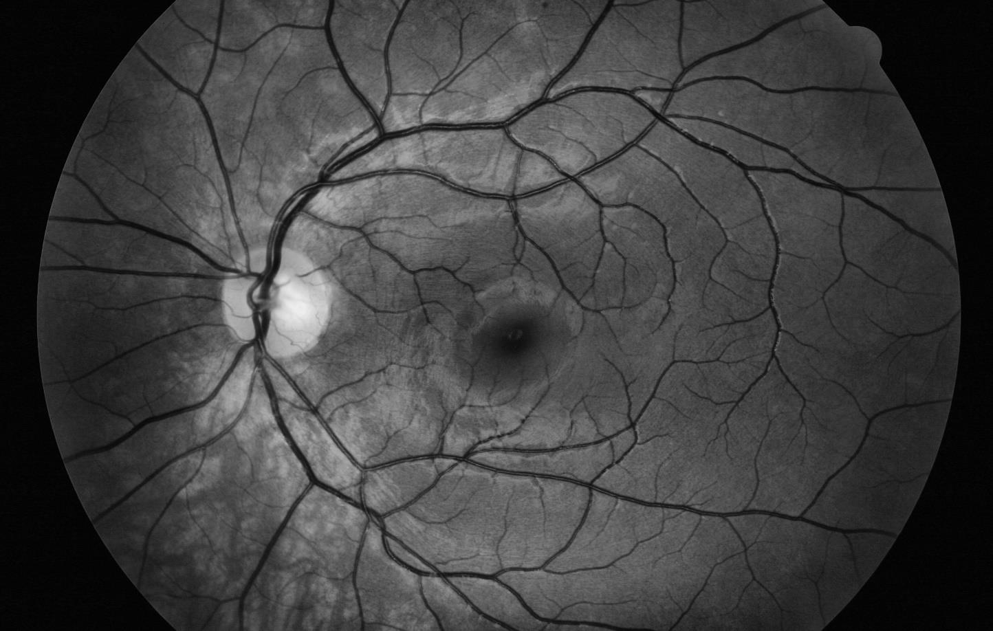
Retinal Blood Supply on OCT After Arterial Occlusion
OCT maps the ischemic territory layer by layer, and in acute occlusion, inner layers become thickened and hyperreflective, while outer layers look relatively normal. Next, this hyperreflectivity fades over several weeks, and finally the inner retina thins, leaving the photoreceptor layers largely preserved.
Deep Plexus Ischemia and PAMM
Paracentral acute middle maculopathy reflects ischemia at the intermediate and deep capillary plexuses, and on OCT, a hyperreflective band appears at the inner nuclear layer. Patients report paracentral scotomas, yet the fundus may look nearly normal. Therefore, PAMM may signal a wider vascular event such as a vein or artery occlusion.
When the Cilioretinal Artery Fails
Isolated cilioretinal occlusion produces a tongue of whitening between the disc and the fovea. It may occur alone, with central retinal vein occlusion, or with giant cell arteritis, and the arteritic form demands urgent systemic evaluation (see cilioretinal artery occlusion).
Ophthalmic Artery Occlusion Pattern
Ophthalmic artery occlusion removes both retinal and choroidal perfusion, and as a result, the retina whitens diffusely, but the cherry-red spot is often absent. Vision frequently falls to no light perception, and OCT also shows hyperreflectivity across both inner and outer layers.
Choroidal Infarction Signs
Choroidal ischemia damages the outer retina and pigment epithelium, and the triangular sign of Amalric reflects a wedge-shaped posterior ciliary territory infarct. In hypertensive crisis, Elschnig spots and Siegrist streaks mark lobular and linear choroidal infarcts.
Imaging Retinal Blood Supply
Fluorescein angiography shows retinal arterial filling and leakage clearly, and choroidal and cilioretinal filling begin before the retinal arterial phase. Indocyanine green angiography displays the choroidal vessels better, and OCT angiography maps capillary plexuses without dye.
Pitfalls to Avoid
- Consider ophthalmic artery occlusion when severe whitening lacks a cherry-red spot
- Look for a cilioretinal artery before predicting visual outcome
- Always exclude giant cell arteritis in older patients with arterial occlusion
- Remember that a near-normal fundus can hide capillary ischemia on OCT


Document what you see
Two smartphone imaging tools built for everyday clinic use — one for the slit lamp, one for the fundus.
From Choroida — the team behind this siteReferences
- Hayreh SS. Acute retinal arterial occlusive disorders. Prog Retin Eye Res. 2011;30:359-394.
- Hayreh SS. Posterior ciliary artery circulation in health and disease: the Weisenfeld lecture. Invest Ophthalmol Vis Sci. 2004;45:749-757.
- Justice J Jr, Lehmann RP. Cilioretinal arteries: a study based on review of stereo fundus photographs and fluorescein angiographic findings. Arch Ophthalmol. 1976;94:1355-1358.
- Sarraf D, Rahimy E, Fawzi AA, et al. Paracentral acute middle maculopathy: a new variant of acute macular neuroretinopathy associated with retinal capillary ischemia. JAMA Ophthalmol. 2013;131:1275-1287.
- Campbell JP, Zhang M, Hwang TS, et al. Detailed vascular anatomy of the human retina by projection-resolved optical coherence tomography angiography. Sci Rep. 2017;7:42201.
Test yourself
A few questions straight from this article.
-
Which circulation supplies the photoreceptors and retinal pigment epithelium?
The retinal circulation serves roughly the inner two thirds of the retina, while the choroid supplies the outer retina, including the photoreceptors and pigment epithelium. -
Where does the central retinal artery enter the optic nerve?
The central retinal artery enters the optic nerve about one centimeter behind the globe, then emerges on the disc and divides into superior and inferior branches. -
Why does a superotemporal branch retinal artery embolus whiten only that quadrant?
The central retinal artery divides into superior and inferior branches, then nasal and temporal arterioles that respect the horizontal raphe, so a branch embolus whitens only its quadrant. -
Which capillary network supplies the thick nerve fibre layer around the optic disc?
Around the disc a radial peripapillary plexus supplies the thick nerve fibre layer, while intermediate and deep plexuses border the inner nuclear layer. -
Which vessels pierce the sclera around the optic nerve to feed the choroid?
Short posterior ciliary arteries pierce the sclera around the optic nerve and feed the choroid, whose fenestrated choriocapillaris has a very high flow rate. -
Which retinal region does a cilioretinal artery usually supply?
A cilioretinal artery arises from the ciliary circulation, hooks round the temporal disc margin and usually supplies part of the papillomacular bundle, so it can preserve central vision in a central occlusion. -
What OCT sign marks paracentral acute middle maculopathy (PAMM)?
PAMM reflects ischemia of the intermediate and deep capillary plexuses and shows a hyperreflective band at the inner nuclear layer, often with a near-normal fundus. -
Which feature helps distinguish ophthalmic artery occlusion from central retinal artery occlusion?
Ophthalmic artery occlusion removes both retinal and choroidal perfusion, so the retina whitens diffusely but the cherry-red spot is often absent and vision often falls to no light perception. -
What fundus pattern does isolated cilioretinal artery occlusion produce?
Isolated cilioretinal occlusion produces a tongue of whitening between the disc and fovea; it may occur alone, with central retinal vein occlusion or with giant cell arteritis. -
What does the triangular sign of Amalric represent?
Choroidal ischemia damages the outer retina and pigment epithelium, and the triangular sign of Amalric reflects a wedge-shaped posterior ciliary territory infarct.

Writes and reviews the clinical articles on Educate. Every post is checked against peer-reviewed sources before it goes live.