Conjunctivitis types share one sign, a red eye, but they differ in discharge, tarsal reaction, nodes and course.
Therefore, a short structured exam usually separates viral, bacterial, allergic, chlamydial and toxic forms. The pattern then decides who needs antibiotics, testing or simple reassurance.
Discharge Across Conjunctivitis Types
First, look at the discharge, because patients describe it well. Watery discharge suggests a viral, allergic or toxic cause, while mucopurulent discharge with lids glued shut in the morning favors bacteria. Hyperpurulent discharge that reaccumulates within minutes after wiping points to gonococcus.
Follicles or Papillae in Conjunctivitis Types
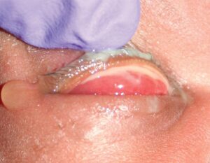
Next, evert the lower lid and examine it at the slit lamp. Follicles are pale, rounded lymphoid nodules with vessels running around their edges, and they suggest viral, chlamydial or toxic disease.
Papillae carry a central vascular core and give a velvety red surface. As a result, papillae favor bacterial or allergic inflammation (see follicles versus papillae).
All-fit Slit-Lamp Adapter
Record and share exactly what you see at the slit lamp. One adapter fits any slit lamp or surgical microscope — and any smartphone.
From Choroida — the team behind this sitePreauricular Nodes
Palpate in front of the tragus in every red eye, because a tender preauricular node strongly supports adenovirus. Chlamydial and gonococcal infections can also enlarge the node. In contrast, allergic, toxic and most simple bacterial cases leave the nodes normal.
Viral Conjunctivitis
Adenovirus usually starts in one eye and reaches the second within days. Then the exam shows watery discharge, follicles, chemosis and sometimes petechial hemorrhages.
Severe cases form pseudomembranes, while subepithelial infiltrates may later blur vision for weeks. Because it spreads easily, hygiene advice matters as much as drops (see viral conjunctivitis).
Bacterial Conjunctivitis
Bacterial disease produces mucopurulent discharge and a papillary reaction, and the onset is acute with the second eye often following. Most cases resolve without treatment within one to two weeks.
However, a Cochrane review found that topical antibiotics modestly speed recovery. Contact lens wearers need closer review because of keratitis risk.
Hyperacute Discharge Needs Same-Day Action
Gonococcal conjunctivitis progresses within hours to marked lid swelling and copious pus, and the node enlarges while the cornea can ulcerate and perforate. Therefore, send a Gram stain and culture, give systemic ceftriaxone and treat sexual contacts. Topical drops alone are never sufficient.
Allergic Conjunctivitis
Itching dominates this picture, and its absence makes allergy unlikely. It involves both eyes, usually with chemosis, stringy mucus and fine papillae, and seasonal timing, atopy and eye rubbing support the diagnosis.
Then treatment relies on dual-action antihistamine drops and allergen avoidance (see allergic conjunctivitis). Giant papillae suggest vernal disease or contact lens irritation instead.
Chlamydial Conjunctivitis
Adult inclusion conjunctivitis is a sexually transmitted infection of young adults that causes a chronic follicular reaction, most prominent in the inferior fornix. Suspect it when presumed viral conjunctivitis lasts beyond two to three weeks. Confirm with nucleic acid amplification testing, then treat with oral azithromycin or doxycycline.
Toxic Conjunctivitis
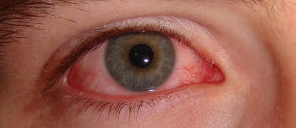
Topical medications and preservatives can inflame the conjunctiva after weeks of use. For example, culprits include aminoglycosides, antivirals, glaucoma drops and benzalkonium chloride, and the reaction may be follicular or papillary, often worst inferiorly.
In addition, inferior punctate keratopathy and a normal node fit this pattern. Finally, symptoms settle after the offending drop stops.
Course and Timing in Conjunctivitis Types
Duration also helps when the exam is equivocal, because viral and bacterial infections usually settle within three weeks. However, chlamydial and toxic cases persist for weeks to months. Therefore, chronic conjunctivitis deserves a fresh history and targeted tests.
Comparing Conjunctivitis Types at a Glance
- Viral: watery discharge, follicles, tender node, one eye then both
- Bacterial: mucopurulent discharge, papillae, usually no node, acute onset
- Gonococcal: hyperpurulent discharge, marked node, rapid corneal risk
- Allergic: itching, stringy mucus, papillae, no node, recurrent
- Chlamydial: follicles, node, mucopurulent discharge, chronic course
- Toxic: watery, follicles or papillae, no node, drop history
Red Flags That Point Away From Conjunctivitis
Conjunctivitis rarely causes reduced vision, photophobia or severe pain, and therefore these symptoms suggest keratitis, uveitis or acute angle closure. Also, look for a corneal infiltrate, ciliary flush and anterior chamber cells. An irregular pupil or raised pressure also changes the diagnosis.
When to Test in Conjunctivitis Types
Most acute cases need no laboratory work. However, culture hyperpurulent discharge, neonatal cases and chronic or recurrent disease, and also request chlamydial testing in chronic follicular disease with sexual risk.
Counseling Across Conjunctivitis Types
Explain that adenovirus spreads by hands and towels, often for up to two weeks, and warn that blur from infiltrates can persist after redness clears. Finally, patients with chlamydia need partner treatment and screening for other infections.
Pitfalls to Avoid
- Do not prescribe antibiotics for every watery red eye
- Never use topical steroids before excluding herpes simplex keratitis
- Do not miss chlamydia in follicular disease lasting over three weeks
- Avoid adding drops when toxicity is the likely cause


Document what you see
Two smartphone imaging tools built for everyday clinic use — one for the slit lamp, one for the fundus.
From Choroida — the team behind this siteReferences
- Azari AA, Barney NP. Conjunctivitis: a systematic review of diagnosis and treatment. JAMA. 2013;310:1721-1729.
- Varu DM, Rhee MK, Akpek EK, et al. Conjunctivitis Preferred Practice Pattern. Ophthalmology. 2019;126:P94-P169.
- Sheikh A, Hurwitz B, van Schayck CP, et al. Antibiotics versus placebo for acute bacterial conjunctivitis. Cochrane Database Syst Rev. 2012;9:CD001211.
- Bowling B. Kanski’s Clinical Ophthalmology: A Systematic Approach. 8th ed. Edinburgh: Elsevier; 2016.
- American Academy of Ophthalmology. Basic and Clinical Science Course, Section 8: External Disease and Cornea. San Francisco: AAO; 2022.
Test yourself
A few questions straight from this article.
-
In conjunctivitis, hyperpurulent discharge that reaccumulates within minutes after wiping points to which organism?
Watery discharge suggests viral, allergic or toxic disease and mucopurulent discharge favours bacteria, but hyperpurulent discharge that reaccumulates within minutes points to gonococcus. -
In conjunctivitis, which feature distinguishes a papilla from a follicle on the everted lid?
Papillae carry a central vascular core and favour bacterial or allergic inflammation, whereas follicles are pale lymphoid nodules with vessels around their edges. -
A red eye with conjunctivitis has a tender preauricular node. Which cause does this strongly support?
A tender preauricular node strongly supports adenovirus; chlamydia and gonococcus can also enlarge it, while allergic, toxic and most simple bacterial cases leave nodes normal. -
Which complication of adenoviral conjunctivitis may blur vision for weeks after the redness clears?
Severe adenoviral cases form pseudomembranes, and subepithelial infiltrates may later blur vision for weeks, even after redness clears. -
Which management is required for gonococcal conjunctivitis?
Gonococcal conjunctivitis can ulcerate and perforate the cornea within hours, so it needs Gram stain, culture, systemic ceftriaxone and contact treatment; topical drops alone are never sufficient. -
Which symptom dominates allergic conjunctivitis, so that its absence makes allergy unlikely?
Itching dominates allergic conjunctivitis, which is bilateral with chemosis, stringy mucus and fine papillae; without itching, allergy is unlikely. -
Presumed viral conjunctivitis persists beyond two to three weeks in a young adult. Which test confirms adult inclusion (chlamydial) conjunctivitis?
Chlamydial conjunctivitis causes a chronic follicular reaction most prominent inferiorly; it is confirmed with nucleic acid amplification testing and treated with oral azithromycin or doxycycline. -
Which preservative is a recognised culprit in toxic conjunctivitis?
Aminoglycosides, antivirals, glaucoma drops and benzalkonium chloride can cause toxic conjunctivitis, with a normal node, inferior punctate keratopathy and relief once the drop stops. -
A red eye with reduced vision, photophobia and severe pain points away from conjunctivitis toward which conditions?
Conjunctivitis rarely causes reduced vision, photophobia or severe pain, so these suggest keratitis, uveitis or acute angle closure; look for infiltrate, ciliary flush and AC cells. -
What is the usual course of bacterial conjunctivitis without treatment?
Most bacterial cases resolve untreated within one to two weeks; a Cochrane review found topical antibiotics only modestly speed recovery.

Writes and reviews the clinical articles on Educate. Every post is checked against peer-reviewed sources before it goes live.