ATN classification is a grading system for myopic maculopathy that scores atrophy, traction and neovascularization separately.
Ruiz-Medrano and colleagues proposed it in 2019 to capture lesions that earlier systems left out. Because each eye receives three scores, one short label describes the whole macula. Therefore, it suits clinics that follow highly myopic patients over many years.
Why a New System Was Needed
The META-PM system grades myopic maculopathy mainly on color fundus photographs, but photographs often miss foveoschisis and early foveal detachment. As a result, an eye with marked traction can still look mild on META-PM. Optical coherence tomography (OCT) shows these traction lesions clearly, so a new framework made sense.
How ATN Classification Differs From META-PM
META-PM uses categories 0 to 4 for atrophy (see the META-PM grading guide), and it then adds plus lesions for lacquer cracks, choroidal neovascularization (CNV) and Fuchs spot. ATN keeps the atrophic categories but adds a separate traction component graded on OCT, and it also moves lacquer cracks into the neovascular component and splits CNV into active and scarred stages.
Fundus Explorer Pro
Photograph the retinal findings described here with the phone already in your pocket — 22 D optics and built-in illumination in one handheld unit.
From Choroida — the team behind this siteImaging Needed for ATN Classification
Color fundus photography grades the atrophic component, and next, OCT grades traction and confirms whether CNV is active. Fluorescein angiography or OCT angiography helps when activity remains uncertain, and fundus autofluorescence also outlines atrophy borders in patchy disease.
Grading Atrophy in ATN Classification
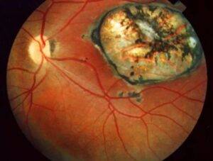
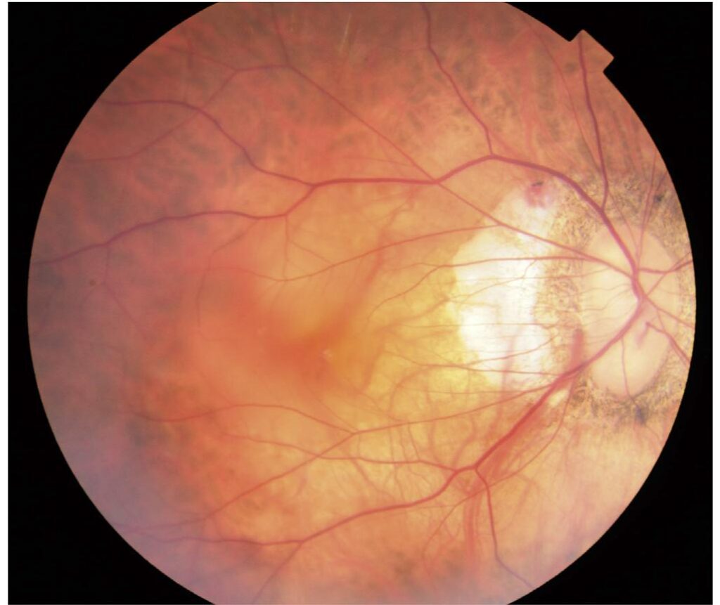
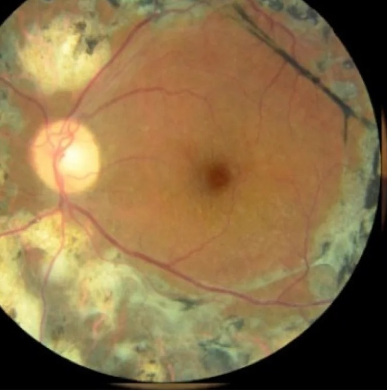
A0 means no myopic retinal lesions, and A1 means a tessellated fundus only. A2 describes diffuse chorioretinal atrophy, a yellowish, ill-defined change around the disc and macula. A3 marks patchy chorioretinal atrophy, where sharply demarcated grayish white areas expose the sclera, and finally A4 means complete macular atrophy.
What the Atrophic Score Means Clinically
Higher A grades predict poorer vision and the damage does not reverse, while patchy atrophy also tends to enlarge slowly over years. Therefore, A3 and A4 eyes need periodic imaging and honest counseling about limited options, and low-vision referral helps once atrophy involves the fovea.
Grading Traction in ATN Classification
T0 means no macular schisis on OCT, and T1 describes inner or outer foveoschisis, while T2 describes both inner and outer foveoschisis. T3 adds a foveal detachment to the schisis, then T4 marks a full-thickness macular hole, and T5 a macular hole with retinal detachment.
How the Traction Score Guides Management
Many T1 and T2 eyes stay stable for long periods, so OCT observation is reasonable, but foveal detachment or falling vision usually prompts surgical referral (see myopic traction maculopathy). Options include vitrectomy with internal limiting membrane peeling, fovea-sparing peeling or macular buckling. Explain that surgery aims to stabilize anatomy, and visual recovery may be partial.
Grading Neovascularization in ATN Classification
N0 means no myopic CNV, and N1 marks macular lacquer cracks, which are linear breaks in Bruch’s membrane. N2a describes active CNV, while N2s describes a scar or Fuchs spot, and lacquer cracks belong here because they often precede CNV.
Why Lacquer Cracks Matter
A patient with a new lacquer crack may notice sudden distortion from a small subretinal hemorrhage, which can occur without any CNV. However, OCT and angiography should exclude CNV before you label it a simple crack bleed.
Treating Active CNV (N2a)
Anti-VEGF injections are first-line therapy for active myopic CNV, and the RADIANCE trial found ranibizumab gave better early visual gains than verteporfin photodynamic therapy. Myopic CNV often needs fewer injections than neovascular age-related macular degeneration, but recurrences occur, so close monitoring after the first injection is wise.
Scarred CNV and Follow-Up
An N2s lesion has no active leakage, so injections offer no benefit, but chorioretinal atrophy often expands around the scar over time. Therefore, follow these eyes for atrophy progression and for reactivation.
Writing an ATN Classification Score
A complete label reads like A3 T1 N2s for one eye, and graders score each component independently, so one eye can carry high and low grades together. In addition, record each eye separately, because the two eyes often differ.
Limits of ATN Classification
The system depends on OCT, which some screening programs lack, and the border between diffuse and patchy atrophy remains partly subjective. Staphyloma and peripapillary changes fall outside its three components, and independent validation is still limited, so many studies report META-PM alongside it.
Counseling Patients About Their Score
Patients need to know which component threatens their vision now. For example, active CNV needs prompt treatment, while early schisis usually needs watching, and teach them to report new distortion or central blur quickly.
Pitfalls to Avoid
- Avoid grading traction from fundus photos alone
- Do not call a crack hemorrhage active CNV without imaging
- Never inject a quiet Fuchs spot
- Remember that a low A grade can coexist with high T or N risk


Document what you see
Two smartphone imaging tools built for everyday clinic use — one for the slit lamp, one for the fundus.
From Choroida — the team behind this siteReferences
- Ruiz-Medrano J, Montero JA, Flores-Moreno I, Arias L, García-Layana A, Ruiz-Moreno JM. Myopic maculopathy: current status and proposal for a new classification and grading system (ATN). Prog Retin Eye Res. 2019;69:80-115.
- Ohno-Matsui K, Kawasaki R, Jonas JB, et al. International photographic classification and grading system for myopic maculopathy. Am J Ophthalmol. 2015;159:877-883.
- Ohno-Matsui K, Wu PC, Yamashiro K, et al. IMI pathologic myopia. Invest Ophthalmol Vis Sci. 2021;62(5):5.
- Wolf S, Balciuniene VJ, Laganovska G, et al. RADIANCE: a randomized controlled study of ranibizumab in patients with choroidal neovascularization secondary to pathologic myopia. Ophthalmology. 2014;121:682-692.
- Shimada N, Tanaka Y, Tokoro T, Ohno-Matsui K. Natural course of myopic traction maculopathy and factors associated with progression or resolution. Am J Ophthalmol. 2013;156:948-957.

Writes and reviews the clinical articles on Educate. Every post is checked against peer-reviewed sources before it goes live.