IOP after cataract surgery often rises within the first hours, even after uneventful phacoemulsification.
Most of these spikes are brief and harmless in a healthy optic nerve, but the same pressure can damage an eye with advanced glaucoma. Therefore, the surgeon must know which rise to watch and which rise to treat.
How Common Is a Rise in IOP After Cataract Surgery?
Transient pressure elevation is one of the most frequent early findings after phacoemulsification, and studies that measured pressure in the first hours found that the peak often occurs several hours after surgery. By the next morning, many eyes have already started to settle. As a result, a routine day-one reading may miss the true peak.
Retained Viscoelastic as a Cause of IOP After Cataract
The ophthalmic viscosurgical device (OVD) protects the endothelium during surgery, but residue left in the eye can obstruct the trabecular meshwork. Material trapped behind the lens is easy to miss, so careful aspiration at the end of the case lowers the risk.
All-fit Slit-Lamp Adapter
Record and share exactly what you see at the slit lamp. One adapter fits any slit lamp or surgical microscope — and any smartphone.
From Choroida — the team behind this siteOther Early Causes
- Postoperative inflammation and fibrin in the anterior chamber
- Retained cortex or nuclear fragments
- Hyphema blocking outflow
- Pigment release, especially in pseudoexfoliation
- Pupillary block from vitreous or an anterior chamber lens
- Aqueous misdirection, which is rare but serious
Which Eyes Are at Risk?
Pre-existing glaucoma and ocular hypertension top the list, while pseudoexfoliation, high axial myopia and known steroid response also raise the risk. Long or complicated surgery increases inflammation and debris, and combined procedures need their own pressure plan (see combined cataract and glaucoma surgery).
Symptoms and Signs of a Spike in IOP After Cataract
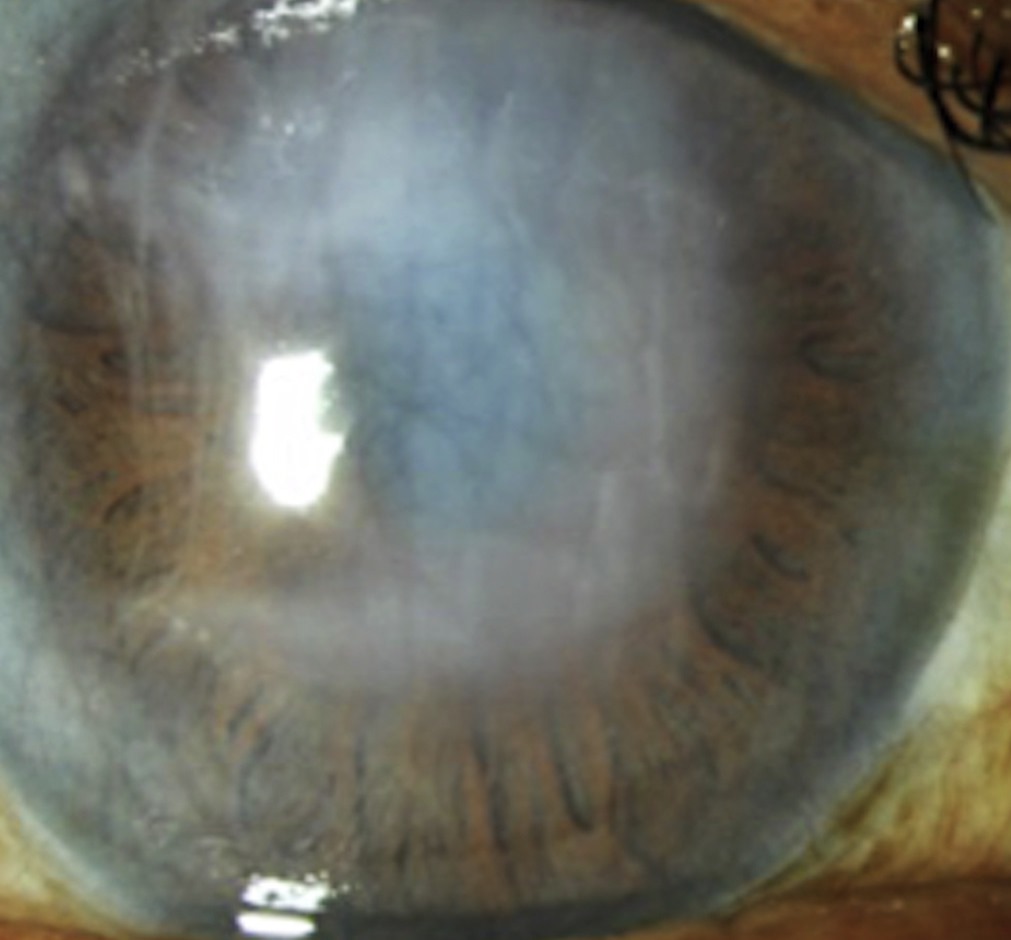
Patients may report brow ache, nausea, halos or blur beyond the expected level, and at the slit lamp, epithelial edema with microcysts suggests a sudden rise. A deep chamber fits retained OVD, while a shallow chamber suggests block or misdirection, and you should also look for blood, fibrin and lens fragments in the angle.
Treatment Thresholds for IOP After Cataract
No single cutoff fits every eye, and in a healthy nerve many surgeons observe pressures in the mid twenties. Many treat readings above about 30 mmHg, or lower values when symptoms appear.
In advanced glaucoma, the threshold falls toward the patient’s usual target. These figures reflect common practice, because trials have not defined a fixed rule.
Medical Treatment of IOP After Cataract
Topical beta-blockers, alpha agonists and carbonic anhydrase inhibitors lower pressure within hours, and oral acetazolamide adds a fast effect for higher spikes. However, check for sulfonamide allergy, kidney disease and electrolyte problems first. Some surgeons avoid prostaglandin analogues in the inflamed early eye, although evidence of harm is limited.
Paracentesis to Release Pressure
Releasing aqueous through the side port gives immediate relief and suits very high pressure with pain and corneal edema. However, the effect is short, and pressure often climbs again. Each entry also carries a small infection risk, so use sterile technique and add medication.
Prevention at the End of Surgery
Thorough OVD removal comes first, and intracameral miotics such as carbachol may blunt the early rise. Some surgeons also give pressure-lowering drops or acetazolamide to high-risk eyes.
Steroid Response and Late IOP After Cataract
A second rise can appear weeks later, during topical steroid therapy, and potent agents such as dexamethasone and prednisolone acetate carry the highest risk. Then, switch to loteprednol or fluorometholone, or taper faster (see steroid-induced glaucoma), and most responses reverse after the steroid stops.
Follow-Up Timing
Randomized data suggest that low-risk, uncomplicated cases may safely skip a day-one visit. However, glaucoma, pseudoexfoliation and complicated cases need a pressure check within 24 hours. Some high-risk eyes benefit from a same-day check several hours after surgery, and next, recheck pressure while the patient still uses steroids.
Complicated Surgery and IOP After Cataract
Capsule rupture brings vitreous, retained fragments and heavier inflammation into the chamber (see vitreous loss), and each of these can raise pressure for days. Therefore, these eyes need closer follow-up and often scheduled pressure-lowering drops, and a dropped nucleus needs prompt vitreoretinal referral.
Counseling the Patient
Explain that a brief pressure rise is common and usually settles, then list warning symptoms: severe pain, nausea, vomiting or worsening blur. Also, ask patients with glaucoma to continue their usual drops unless told otherwise, and finally, give a clear number to call after hours.
Pitfalls to Avoid
- Do not rely on a day-one reading in a high-risk eye
- Never ignore nausea and brow ache as simple postoperative discomfort
- Do not forget steroid response at the one-month visit
- Avoid repeated paracentesis without adding medical treatment


Document what you see
Two smartphone imaging tools built for everyday clinic use — one for the slit lamp, one for the fundus.
From Choroida — the team behind this siteReferences
- Shingleton BJ, Wadhwani RA, O’Donoghue MW, Baylus S, Hoey H. Evaluation of intraocular pressure in the immediate period after phacoemulsification. J Cataract Refract Surg. 2001;27:524-527.
- Grzybowski A, Kanclerz P. Early postoperative intraocular pressure elevation following cataract surgery. Curr Opin Ophthalmol. 2019;30:56-62.
- Tinley CG, Frost A, Hakin KN, McDermott W, Ewings P. Is visual outcome compromised when next day review is omitted after phacoemulsification surgery? A randomised control trial. Br J Ophthalmol. 2003;87:1350-1355.
- Olson RJ, Braga-Mele R, Chen SH, et al. Cataract in the Adult Eye Preferred Practice Pattern. Ophthalmology. 2017;124:P1-P119.
- American Academy of Ophthalmology. Basic and Clinical Science Course, Section 11: Lens and Cataract. San Francisco: AAO; 2023.
Test yourself
A few questions straight from this article.
-
When does the intraocular pressure peak often occur after uneventful phacoemulsification?
Studies measuring pressure in the first hours found the peak often occurs several hours after surgery, and many eyes are settling by morning, so a day-one reading may miss it. -
How does retained ophthalmic viscosurgical device (OVD) raise intraocular pressure after cataract surgery?
OVD protects the endothelium during surgery, but residue, including material trapped behind the lens, can obstruct the trabecular meshwork; careful aspiration lowers the risk. -
Which eyes carry the highest risk of a pressure rise after cataract surgery?
Pre-existing glaucoma and ocular hypertension top the list, with pseudoexfoliation, high axial myopia, known steroid response and long or complicated surgery also raising risk. -
After cataract surgery with a pressure spike, what does a shallow anterior chamber suggest?
A deep chamber fits retained OVD, while a shallow chamber suggests block or misdirection; the angle should also be checked for blood, fibrin and lens fragments. -
In an eye with a healthy optic nerve, above roughly what pressure do many surgeons treat a post-cataract spike?
Many surgeons observe mid-twenties pressures in a healthy nerve and treat above about 30 mmHg or lower when symptomatic; in advanced glaucoma the threshold falls toward the usual target. -
Before giving oral acetazolamide for a post-cataract pressure spike, what should be checked?
Oral acetazolamide adds a fast effect for higher spikes, but sulfonamide allergy, kidney disease and electrolyte problems must be checked first. -
What is the main limitation of anterior chamber paracentesis for a post-cataract pressure spike?
Releasing aqueous via the side port gives immediate relief, but the effect is short and each entry carries a small infection risk, so medication should be added. -
Which intracameral agent given at the end of cataract surgery may blunt the early pressure rise?
Thorough OVD removal comes first, and intracameral miotics such as carbachol may blunt the early rise. -
A pressure rise appears weeks after cataract surgery during dexamethasone drops. What change is advised?
Potent steroids such as dexamethasone and prednisolone acetate carry the highest risk; switching to loteprednol or fluorometholone or tapering faster helps, and most responses reverse after stopping. -
Which patients need a pressure check within 24 hours of cataract surgery rather than skipping the day-one visit?
Randomized data suggest low-risk uncomplicated cases may skip day one, but glaucoma, pseudoexfoliation and complicated cases need a check within 24 hours.

Writes and reviews the clinical articles on Educate. Every post is checked against peer-reviewed sources before it goes live.