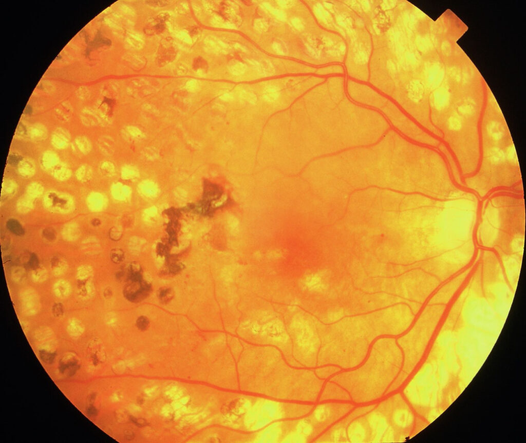
Retinal laser scars are the permanent chorioretinal marks left after thermal photocoagulation of the retina.
They follow panretinal photocoagulation (PRP), focal or grid macular laser, and laser retinopexy, and each treatment leaves a typical pattern, so the fundus often reveals the patient’s history. However, some inflammatory and degenerative scars look similar, and the distinction changes management.
How Retinal Laser Scars Form
Melanin in the retinal pigment epithelium (RPE) and choroid absorbs most of the laser energy, and heat then spreads to the photoreceptors and, with stronger burns, to the inner retina. As a result, the outer retina whitens within seconds of treatment, and over the following weeks the RPE proliferates and migrates, while photoreceptors inside the burn die.
The Fresh Burn Versus the Mature Scar
A fresh burn looks gray-white and slightly opaque with soft edges, and within a few weeks pigment begins to gather at the border or across the spot. Eventually, a mature scar shows a pale atrophic center, a dark pigment rim, or a mottled mix of both, so the color gives a rough idea of how old the treatment is.
Fundus Explorer Pro
Photograph the retinal findings described here with the phone already in your pocket — 22 D optics and built-in illumination in one handheld unit.
From Choroida — the team behind this siteRetinal Laser Scars After PRP
PRP scars are numerous, round and similar in size, and they fill the midperiphery and periphery, while the posterior pole and disc margin stay untreated. Conventional PRP usually places burns about one burn width apart, so the scars form loose rows between vessels.
Patterned scanning lasers such as PASCAL, by contrast, leave lighter scars in neat square or arc grids. In addition, heavy PRP can leave confluent atrophic areas where bare choroidal vessels show through.
Focal and Grid Macular Scars
Focal laser for diabetic macular edema targets leaking microaneurysms with small burns, while grid laser places light burns across thickened retina and spares the foveal avascular zone. As a result, the scars appear as tiny pigmented or atrophic dots scattered around the fovea, and many modern light burns look subtle on color photographs but clearer on autofluorescence.
Retinopexy Scars Around a Break
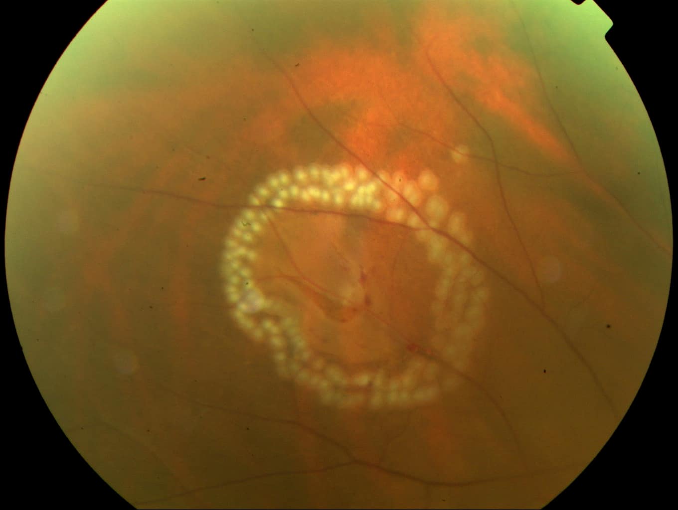
Laser retinopexy surrounds a tear or hole with two or three rows of confluent burns. The mature result is a pigmented ring or horseshoe that encircles the break (see laser retinopexy).
Therefore, a peripheral pigment ring with a break or flap at its center points strongly to prior treatment. Check that the ring is complete, because an anterior gap can still allow fluid to pass.
Retinal Laser Scars on OCT
OCT across a fresh burn shows a hyperreflective band in the outer retina. Later, the outer nuclear layer thins, and the ellipsoid zone and RPE lose continuity within the scar, while the choroid behind the scar often shows increased signal transmission. Hyperreflective clumps above the RPE usually represent migrated pigment (see ellipsoid zone disruption).
Autofluorescence in Retinal Laser Scars
Fundus autofluorescence shows mature scars as dark spots because the RPE is lost, and some scars also carry a bright rim where the RPE is stressed or hyperplastic. Fresh burns may look different, so interpret the image alongside the treatment date.
Scar Expansion Over Time
Laser scars can change, especially in the macula, and Schatz and colleagues described progressive enlargement of scars after grid laser for diffuse diabetic macular edema. Over years, atrophy can spread and merge, and scars near the fovea may threaten central vision. Therefore, document macular scars with photographs or FAF and compare them at follow-up.
Complications Seen at the Scar Edge
Choroidal neovascularization can occasionally develop at the edge of a laser scar, particularly after heavy burns. Look for new subretinal fluid, hemorrhage or a gray membrane next to an old scar, and OCT angiography or fluorescein angiography then helps confirm the diagnosis.
Telling Retinal Laser Scars From Chorioretinitis
Laser scars are uniform in size, regularly spaced and arranged in patterns, while chorioretinitis scars, however, vary in size and shape and cluster without geometry. Toxoplasmosis typically leaves a larger punched-out, pigmented scar, often with satellite lesions (see toxoplasma scars). Fluffy whitening at the edge of an old scar suggests reactivation, which laser scars never show.
Other Mimics to Exclude
- Paving stone degeneration: round, pale peripheral patches with pigmented borders near the ora
- Presumed ocular histoplasmosis: small punched-out spots plus peripapillary atrophy
- Multifocal choroiditis and punctate inner choroidopathy: irregular scars, often in myopic eyes
- Cryotherapy scars: larger, heavily pigmented and less sharply defined
Counseling Patients About Retinal Laser Scars
Explain that the scars are expected and show that the treatment reached its target, but warn that extensive PRP can reduce peripheral and night vision. Scars need no treatment, and they do not disappear, and finally, ask patients to report new distortion or central blur promptly.
Pitfalls to Avoid
- Do not label regular peripheral rows as old inflammation before considering prior PRP
- Never ignore fresh whitening at a scar edge
- Do not assume a retinopexy ring is complete without indirect ophthalmoscopy


Document what you see
Two smartphone imaging tools built for everyday clinic use — one for the slit lamp, one for the fundus.
From Choroida — the team behind this siteReferences
- Diabetic Retinopathy Study Research Group. Photocoagulation treatment of proliferative diabetic retinopathy: DRS report number 8. Ophthalmology. 1981;88:583-600.
- Early Treatment Diabetic Retinopathy Study Research Group. Photocoagulation for diabetic macular edema: ETDRS report number 1. Arch Ophthalmol. 1985;103:1796-1806.
- Schatz H, Madeira D, McDonald HR, Johnson RN. Progressive enlargement of laser scars following grid laser photocoagulation for diffuse diabetic macular edema. Arch Ophthalmol. 1991;109:1549-1551.
- Bowling B. Kanski’s Clinical Ophthalmology: A Systematic Approach. 8th ed. Elsevier; 2016.
Test yourself
A few questions straight from this article.
-
In retinal laser photocoagulation, what absorbs most of the laser energy that creates the scar?
Melanin in the retinal pigment epithelium and choroid absorbs most of the energy; heat then spreads to the photoreceptors and, with stronger burns, to the inner retina. -
How does a fresh retinal laser burn typically look on fundus examination?
A fresh burn is gray-white with soft edges; pigment gathers over weeks, and a mature scar shows a pale atrophic centre, a dark pigment rim, or both. -
Where are panretinal photocoagulation (PRP) scars distributed in the fundus?
PRP scars are numerous, round and similar in size, filling the midperiphery and periphery while the posterior pole and disc margin stay untreated. -
How far apart does conventional panretinal photocoagulation usually place its burns?
Conventional PRP places burns about one burn width apart, so the scars form loose rows between the vessels. -
What scar pattern do patterned scanning lasers such as PASCAL leave in the retina?
Unlike conventional PRP rows, patterned scanning lasers such as PASCAL leave lighter scars arranged in neat square or arc grids. -
How does laser retinopexy treat a peripheral retinal tear or hole?
Retinopexy surrounds the break with two or three rows of confluent burns, maturing into a pigmented ring; the ring should be complete because an anterior gap can still let fluid pass. -
How do mature retinal laser scars appear on fundus autofluorescence?
Mature scars are dark on autofluorescence because the RPE is lost; some carry a bright rim where the RPE is stressed or hyperplastic. -
Which complication can develop at the edge of a retinal laser scar, especially after heavy burns?
Choroidal neovascularization can occasionally arise at a scar edge; new subretinal fluid, haemorrhage or a gray membrane next to an old scar should prompt OCT angiography or fluorescein angiography. -
Which feature helps distinguish retinal laser scars from chorioretinitis scars?
Laser scars are uniform and regularly spaced in patterns, whereas chorioretinitis scars vary in size and shape and cluster without geometry. -
Why should macular laser scars be photographed or imaged with autofluorescence and compared at follow-up?
Progressive enlargement of scars has been described after grid laser for diffuse diabetic macular edema; atrophy can spread and merge over years and threaten central vision.

Writes and reviews the clinical articles on Educate. Every post is checked against peer-reviewed sources before it goes live.