Snap back test results show how well the lower eyelid holds its position against the globe.
The test takes seconds at the slit lamp or chairside.
Together with the distraction test, it grades horizontal lid laxity, the main driver of involutional ectropion.
As a result, these two bedside checks also shape surgical planning.
What the Snap Back Test Measures
The snap back test assesses the elastic recoil of the lower lid.
A healthy tarsus and firm canthal tendons return the lid to the globe almost instantly.
With age, however, the tendons stretch and the orbicularis loses tone.
Therefore, the lid returns slowly or stays away from the eye.
How to Perform the Snap Back Test
First, ask the patient to look straight ahead and avoid blinking.
Next, pull the lower lid down and away from the globe with a fingertip.
Then release it and watch the lid margin.
Finally, note how long the lid takes to meet the globe, and whether a blink helps it return.
All-fit Slit-Lamp Adapter
Record and share exactly what you see at the slit lamp. One adapter fits any slit lamp or surgical microscope — and any smartphone.
From Choroida — the team behind this siteGrading the Snap Back Test
Several scales exist, and none is universally standard.
For example, one commonly cited approach grades recoil from 0 to 4:
- Immediate return to the globe: grade 0
- Slow return within 2 to 3 seconds: grade 1
- Return within 4 to 5 seconds: grade 2
- Delayed return beyond 5 seconds, only after a blink: grade 3
- No return even after blinking: grade 4
In practice, use the same method at each visit.
The Distraction Test
The distraction test measures horizontal laxity in millimeters.
First, pinch the central lower lid and pull it directly forward, away from the cornea.
Then measure the gap between the lid margin and the corneal surface.
Most texts consider more than 6 mm abnormal, although some surgeons use 8 mm.
In addition, a lid that distracts easily often fails the snap back test too.
Snap Back Test Versus Distraction Test
The two tests answer related questions.
Distraction shows how far the lid can travel, so it mainly reflects tendon length.
Recoil, by contrast, depends on both tendon tension and orbicularis tone.
Therefore, a lid can distract moderately yet recoil poorly in a patient with facial weakness.
Checking the Canthal Tendons
Laxity is rarely uniform, so test each canthus separately.
For the medial canthal tendon, pull the lower lid laterally and watch the punctum.
Displacement of several millimeters, or toward the limbus, suggests significant medial laxity.
Similarly, a rounded and mobile lateral canthus on medial traction points to lateral tendon laxity.
Snap Back Test in Involutional Ectropion
Horizontal laxity is the main abnormality in involutional ectropion.
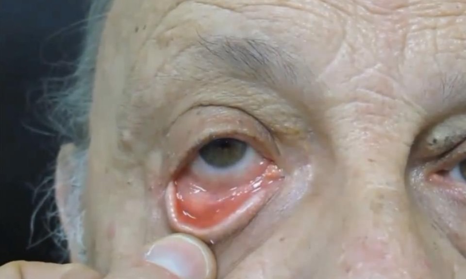
Typically, a poor snap back test and wide distraction accompany a sagging, everted lid (see ectropion and its effects).
In addition, lower lid retractor disinsertion may coexist and needs separate repair.
Look for a deep inferior fornix and reduced lid excursion on downgaze.
Excluding Other Causes of Ectropion
A lax lid can still hide a second mechanism.
Cicatricial ectropion follows skin shortage from sun damage, scars or dermatitis.
Ask the patient to look up and open the mouth, because increased eversion suggests a skin deficit.
Also push the lid up with a finger; if it cannot reach its normal position, the skin is short.
How Laxity Guides Surgery
When horizontal laxity dominates, horizontal shortening is usually the first step.
The lateral tarsal strip shortens the lid and reanchors it to the lateral orbital rim.
Medial canthal laxity, however, may need plication or a separate medial procedure.
Otherwise, lateral tightening can drag the punctum away from the tear lake.
Snap Back Test Before Blepharoplasty
Lower lid blepharoplasty can unmask hidden laxity.
As a result, retraction or ectropion may follow skin or muscle removal.
Therefore, perform both tests before every lower lid procedure.
A poor result often prompts a concurrent canthal tightening.
Snap Back Test in Facial Palsy
In facial nerve palsy, orbicularis weakness slows recoil independent of tendon length.
Therefore, interpret a slow result with caution.
Exposure keratopathy may coexist, so stain the cornea with fluorescein (see slit lamp exam).
Management then focuses on corneal protection alongside any lid tightening.
Counseling the Patient
Explain that the lid has stretched with age, much like a loose elastic band.
Then show the patient how slowly the lid returns after release.
Also explain that tearing may improve, while blocked drainage can need separate treatment.
Pitfalls to Avoid
- Do not let the patient blink before the lid recoils
- Avoid testing a supine or sedated patient, because tone changes
- Do not skip separate canthal tendon assessment
- Never blame laxity alone before checking for skin shortage
- Record the method so follow-up results stay comparable


Document what you see
Two smartphone imaging tools built for everyday clinic use — one for the slit lamp, one for the fundus.
From Choroida — the team behind this siteReferences
- Anderson RL, Gordy DD. The tarsal strip procedure. Arch Ophthalmol. 1979;97:2192-2196.
- Jordan DR, Anderson RL. The lateral tarsal strip revisited: the enhanced tarsal strip. Arch Ophthalmol. 1989;107:604-606.
- American Academy of Ophthalmology. Basic and Clinical Science Course, Section 7: Oculofacial Plastic and Orbital Surgery. San Francisco: AAO; 2023.
- Salmon JF. Kanski’s Clinical Ophthalmology: A Systematic Approach. 9th ed. Edinburgh: Elsevier; 2020.

Writes and reviews the clinical articles on Educate. Every post is checked against peer-reviewed sources before it goes live.