Best disease stages describe the evolution of an autosomal dominant macular dystrophy caused by BEST1 variants.
Clinicians usually follow the Gass sequence, although numbering differs slightly between sources. Because lesions may skip or repeat a phase, the stage describes appearance and does not fix a timeline.
How Best Disease Stages Are Organized
Most schemes begin with a previtelliform phase and then move through vitelliform, pseudohypopyon and vitelliruptive appearances. Atrophy follows in many eyes, and some develop fibrosis or choroidal neovascularization (CNV). Because the two eyes may sit at different stages in one patient, document each eye separately.
Why Material Accumulates
BEST1 encodes bestrophin-1, a protein in the basolateral membrane of the retinal pigment epithelium (RPE). Faulty ion transport probably disturbs fluid handling and outer segment clearance at the RPE surface. As a result, yellow material collects in the subretinal space.
Fundus Explorer Pro
Photograph the retinal findings described here with the phone already in your pocket — 22 D optics and built-in illumination in one handheld unit.
From Choroida — the team behind this sitePrevitelliform Stage
The macula looks normal or shows only subtle RPE mottling, and vision is usually normal, so family screening often detects this stage. However, the electrooculogram (EOG) is typically abnormal already.
Vitelliform Stage
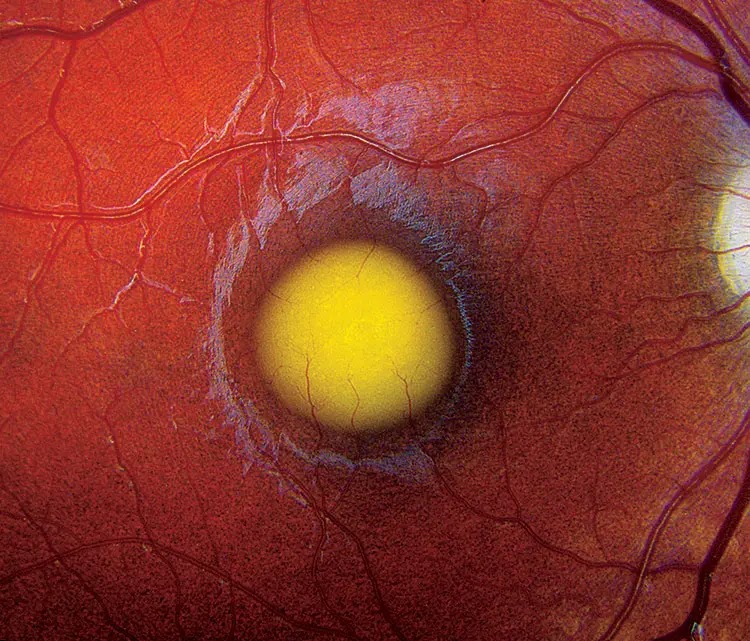
Here the examiner sees the classic round, yellow, egg yolk lesion at the fovea. It commonly appears in childhood, and the patient may report few symptoms despite its striking look.
On OCT, hyperreflective material lies between the RPE and the photoreceptor outer segments. Many patients keep good acuity at this stage because the photoreceptors remain relatively intact.
Pseudohypopyon Stage
Next, the material may separate and settle inferiorly under gravity, forming a yellow fluid level. OCT shows dense material below and a less reflective space above it.
Vitelliruptive Stage
The yolk then breaks up, producing the familiar “scrambled egg” pattern. Patients may now notice more blur, while acuity varies widely between eyes. OCT typically shows a mixed picture of residual hyperreflective deposits and an optically empty space, and the outer retinal bands may look irregular over the lesion.
Reading OCT Fluid in Best Disease Stages
That empty space often looks like active exudation, but it may persist for long periods without CNV. Therefore, fluid alone cannot support anti-VEGF injections.
Look for sudden symptoms, hemorrhage, a subretinal hyperreflective lesion or a vascular network. OCT angiography can then help confirm or exclude a neovascular membrane.
Atrophic Stage
Eventually the yellow material fades and leaves a pale area of macular atrophy. OCT shows thinning of the outer retina and RPE, often with loss of the ellipsoid zone.
Because photoreceptors are now lost, central vision usually declines more clearly at this stage. Assessing ellipsoid zone disruption also helps explain the acuity you measure.
Fibrotic and Neovascular Stages
Some eyes develop a white subretinal scar, sometimes with a raised central fibrotic mound. CNV can occur too, including in children and young adults, and patients with CNV may report abrupt distortion or a fresh drop in vision.
Autofluorescence in Best Disease Stages
Vitelliform material appears intensely bright on fundus autofluorescence. During the vitelliruptive stage, the signal becomes patchy as material redistributes.
Atrophic areas then appear dark because the RPE fluorophores are lost. Consequently, autofluorescence helps track change between visits without any dye.
EOG and ERG Testing
The EOG reflects RPE function, and the Arden ratio is typically reduced, often below 1.5. Meanwhile, the full-field ERG usually remains normal, since the disease is not a generalized photoreceptor dystrophy.
A normal EOG should prompt a review of the diagnosis. Genetic testing can then confirm a BEST1 variant.
Conditions to Exclude
Several disorders can mimic these appearances, especially in adults. For example, adult-onset foveomacular vitelliform dystrophy usually forms smaller lesions, and the EOG is often normal. Pattern dystrophy, central serous chorioretinopathy and vitelliform lesions with drusen also enter the differential.
Managing Best Disease Stages
No treatment currently reverses the underlying defect or clears the vitelliform material. Therefore, management centers on monitoring, refraction, low vision support and treating complications.
Anti-VEGF therapy can help when CNV is confirmed, although the evidence comes mainly from small series. Injections cannot restore photoreceptors already lost to atrophy, and patients should hear that before treatment.
Counseling Families on Best Disease Stages
Each child of an affected parent has a 50% chance of inheriting the variant. However, expressivity varies, and some carriers show few visible fundus changes. Explain that a dramatic lesion does not automatically mean imminent blindness.
Pitfalls to Avoid
- Do not treat persistent subretinal space as CNV without supporting signs
- Never exclude Best disease because the fundus looks normal in a relative
- Do not predict vision from the stage alone
- Avoid labeling every adult vitelliform lesion as Best disease


Document what you see
Two smartphone imaging tools built for everyday clinic use — one for the slit lamp, one for the fundus.
From Choroida — the team behind this siteReferences
- Boon CJ, Klevering BJ, Leroy BP, et al. The spectrum of ocular phenotypes caused by mutations in the BEST1 gene. Prog Retin Eye Res. 2009;28:187-205.
- Spaide RF, Noble K, Morgan A, Freund KB. Vitelliform macular dystrophy. Ophthalmology. 2006;113:1392-1400.
- Ferrara DC, Costa RA, Tsang S, et al. Multimodal fundus imaging in Best vitelliform macular dystrophy. Graefes Arch Clin Exp Ophthalmol. 2010;248:1377-1385.
- Booij JC, Boon CJ, van Schooneveld MJ, et al. Course of visual decline in relation to the Best1 genotype in vitelliform macular dystrophy. Ophthalmology. 2010;117:1415-1422.
Test yourself
A few questions straight from this article.
-
In autosomal dominant Best disease, what is the chance that each child of an affected parent inherits the variant?
Each child of an affected parent has a 50% chance of inheriting the BEST1 variant, although expressivity varies and some carriers show few fundus changes. -
Where is bestrophin-1, the protein affected in Best disease, located?
BEST1 encodes bestrophin-1 in the basolateral RPE membrane; faulty ion transport probably disturbs fluid handling and outer segment clearance, so yellow material collects subretinally. -
How is the previtelliform stage of Best disease often detected?
In the previtelliform stage the macula looks normal or shows subtle RPE mottling and vision is usually normal, so family screening often detects it, even though the EOG is already abnormal. -
On OCT in the vitelliform stage of Best disease, where does the hyperreflective material lie?
OCT shows hyperreflective material between the RPE and photoreceptor outer segments, and many patients keep good acuity because the photoreceptors remain relatively intact. -
What defines the pseudohypopyon stage of Best disease?
In the pseudohypopyon stage the material separates and settles inferiorly under gravity, forming a yellow fluid level, with dense material below and a less reflective space above on OCT. -
In Best disease, does an optically empty subretinal space on OCT alone justify anti-VEGF injections?
The empty space can look like exudation yet persist without CNV, so fluid alone cannot support injections; sudden symptoms, haemorrhage, a hyperreflective lesion or a vascular network on OCTA are sought instead. -
At which stage of Best disease does central vision usually decline more clearly?
In the atrophic stage the material fades, leaving outer retinal and RPE thinning with ellipsoid zone loss; with photoreceptors lost, central vision usually declines more clearly. -
How does vitelliform material appear on fundus autofluorescence in Best disease?
Vitelliform material is intensely bright on autofluorescence, becomes patchy in the vitelliruptive stage, and atrophic areas appear dark as RPE fluorophores are lost. -
Which electrophysiology pattern is typical of Best disease?
The EOG reflects RPE function and its Arden ratio is typically below 1.5, while the full-field ERG usually stays normal because Best disease is not a generalized photoreceptor dystrophy. -
How does adult-onset foveomacular vitelliform dystrophy usually differ from Best disease?
Adult-onset foveomacular vitelliform dystrophy usually forms smaller lesions with an often normal EOG, and a normal EOG should prompt review of a Best disease diagnosis.

Writes and reviews the clinical articles on Educate. Every post is checked against peer-reviewed sources before it goes live.