ROP classification describes each eye using zone, stage, extent and plus disease.
The current system, ICROP3, appeared in 2021 and updated the earlier international reports.
Because these terms guide follow-up and treatment, examiners should apply them consistently.
For the wider workup, see our ROP diagnosis guide.
What ROP Classification Records
Retinal vessels grow from the optic disc toward the ora serrata until around term.
Preterm birth disturbs this growth, so the examiner maps where the vessels stop.
Zone then records location, while stage describes the abnormal response at the vascular junction.
How ROP Classification Evolved
The 1984 report introduced zones, stages, extent and plus disease.
Next, the 2005 revision added preplus disease and aggressive posterior ROP.
Finally, ICROP3 added posterior zone II, the notch, stage 5 subtypes and reactivation terminology.
Fundus Explorer Pro
Photograph the retinal findings described here with the phone already in your pocket — 22 D optics and built-in illumination in one handheld unit.
From Choroida — the team behind this siteZones in ROP Classification
All zones center on the optic disc because vascular growth starts there.
Zone I is a circle whose radius equals twice the disc-to-fovea distance.
Zone II extends from that border to a circle reaching the nasal ora serrata.
The remaining temporal crescent forms zone III.
Posterior Zone II and the Notch
ICROP3 defines posterior zone II as a band two disc diameters beyond the zone I border.
Examiners watch this band closely, because disease here may behave more severely than peripheral zone II disease.
A notch is a one to two clock-hour incursion of the lesion along the horizontal meridian.
For example, you would record “zone I secondary to notch” when it reaches zone I.
Stages 1 to 3 in ROP Classification
Stage 1 is a thin, flat, white demarcation line between vascular and avascular retina.
Stage 2 follows when that line gains height and width, forming a ridge.
Then, stage 3 adds extraretinal neovascular proliferation or flat neovascularization.
Stages 4 and 5: Retinal Detachment
Stage 4 means partial detachment, with 4A sparing the fovea and 4B involving it.
Stage 5 means total detachment, and ICROP3 divides it into three subtypes.
In 5A, ophthalmoscopy still shows the disc, which suggests an open funnel.
In 5B, retrolental fibrovascular tissue or a closed funnel hides the disc.
Finally, 5C adds anterior segment changes, such as lens displacement and marked chamber shallowing.
Extent in Clock Hours
Extent records the involved retina in clock hours.
CRYO-ROP threshold disease required five contiguous or eight cumulative hours of stage 3 with plus disease in zone I or II.
However, ETROP criteria later moved decisions toward zone, stage and plus status.
Plus Disease in ROP Classification
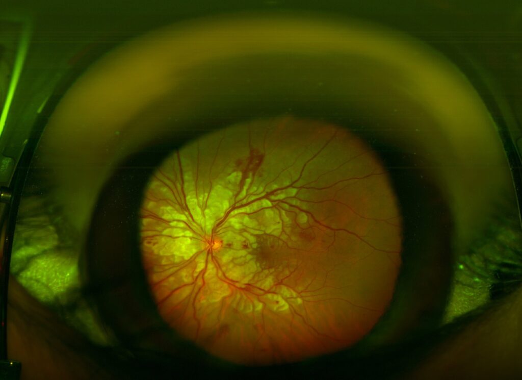
Plus disease means abnormal dilation and tortuosity of posterior pole vessels within zone I.
ICROP3 no longer relies on counting involved quadrants.
Instead, it treats vascular change as a continuum from normal through preplus to plus.
Preplus therefore describes abnormal vessels that fall short of plus disease.
Aggressive ROP
ICROP3 replaced aggressive posterior ROP with aggressive ROP (A-ROP), because this pattern can appear beyond the posterior retina.
The examiner sees rapid pathologic neovascularization and severe plus disease, often without typical stage progression.
See also our review of aggressive posterior ROP.
Regression, Reactivation and Avascular Retina
Regression may occur spontaneously or after treatment, so record whether it is complete.
Persistent avascular retina describes peripheral retina left unvascularized, and its location belongs in the notes.
Reactivation means new ROP features after regression, particularly following anti-VEGF therapy.
Documenting ROP Classification
Record each eye separately, using the most posterior zone and the most severe stage.
Then add extent, plus status and any regression.
For example: right eye, zone II, stage 3, four clock hours, preplus disease.
From ROP Classification to Treatment
ETROP type 1 includes zone I disease with plus, zone I stage 3, and zone II stage 2 or 3 with plus.
Type 2 includes zone I stage 1 or 2 without plus, and zone II stage 3 without plus.
ETROP supported treating type 1 eyes and closely observing type 2 eyes.
Treatment choice, however, sits outside the classification (see ROP treatment options).
Explaining ROP Classification to Parents
Describe zone as how far the vessels have grown.
Next, describe stage as the severity of changes at the vessel edge.
Explain that disease can change quickly, so a mild finding today does not remove the need for the next examination.
Pitfalls to Avoid
- Do not overlook a notch when assigning the zone
- Never diagnose plus disease from vessels outside zone I
- Do not assume A-ROP occurs only in posterior retina
- Do not stop follow-up at first regression after anti-VEGF


Document what you see
Two smartphone imaging tools built for everyday clinic use — one for the slit lamp, one for the fundus.
From Choroida — the team behind this siteReferences
- Chiang MF, Quinn GE, Fielder AR, et al. International Classification of Retinopathy of Prematurity, Third Edition. Ophthalmology. 2021;128:e51-e68.
- International Committee for the Classification of Retinopathy of Prematurity. The International Classification of Retinopathy of Prematurity revisited. Arch Ophthalmol. 2005;123:991-999.
- Committee for the Classification of Retinopathy of Prematurity. An international classification of retinopathy of prematurity. Arch Ophthalmol. 1984;102:1130-1134.
- Early Treatment for Retinopathy of Prematurity Cooperative Group. Revised indications for the treatment of retinopathy of prematurity. Arch Ophthalmol. 2003;121:1684-1694.
- Cryotherapy for Retinopathy of Prematurity Cooperative Group. Multicenter trial of cryotherapy for retinopathy of prematurity: preliminary results. Arch Ophthalmol. 1988;106:471-479.
Test yourself
A few questions straight from this article.
-
In ROP classification, how is the radius of zone I defined?
All zones center on the optic disc because vascular growth starts there, and zone I is a circle whose radius equals twice the disc-to-fovea distance. -
How does ICROP3 define posterior zone II in retinopathy of prematurity?
ICROP3 defines posterior zone II as a band two disc diameters beyond the zone I border, and disease here may behave more severely than peripheral zone II disease. -
What is a notch in the ICROP3 classification of retinopathy of prematurity?
A notch is a one to two clock-hour incursion along the horizontal meridian, recorded for example as zone I secondary to notch when it reaches zone I. -
Which stage of retinopathy of prematurity forms when the demarcation line gains height and width?
Stage 1 is a thin, flat, white demarcation line, and stage 2 follows when that line gains height and width to form a ridge. -
Which ICROP3 subtype of stage 5 retinopathy of prematurity has retrolental tissue or a closed funnel hiding the disc?
In 5A the disc is still visible, in 5B retrolental fibrovascular tissue or a closed funnel hides it, and 5C adds anterior segment changes such as lens displacement and chamber shallowing. -
What extent of stage 3 with plus disease in zone I or II defined CRYO-ROP threshold disease?
CRYO-ROP threshold disease required five contiguous or eight cumulative clock hours of stage 3 with plus disease in zone I or II, though ETROP later moved decisions toward zone, stage and plus status. -
How does ICROP3 approach plus disease in retinopathy of prematurity?
Plus disease means abnormal dilation and tortuosity of posterior pole vessels within zone I, and ICROP3 no longer relies on counting quadrants but treats the change as a continuum. -
Why did ICROP3 replace aggressive posterior ROP with the term aggressive ROP?
ICROP3 made the change because this pattern can appear beyond the posterior retina; it shows rapid pathologic neovascularization and severe plus disease, often without typical stage progression. -
What does reactivation mean in ICROP3 terminology for retinopathy of prematurity?
Reactivation means new ROP features after regression, particularly following anti-VEGF therapy, so follow-up should not stop at first regression. -
Which eye meets ETROP type 1 criteria in retinopathy of prematurity?
Type 1 includes zone I disease with plus, zone I stage 3, and zone II stage 2 or 3 with plus; ETROP supported treating type 1 eyes and closely observing type 2 eyes.

Writes and reviews the clinical articles on Educate. Every post is checked against peer-reviewed sources before it goes live.