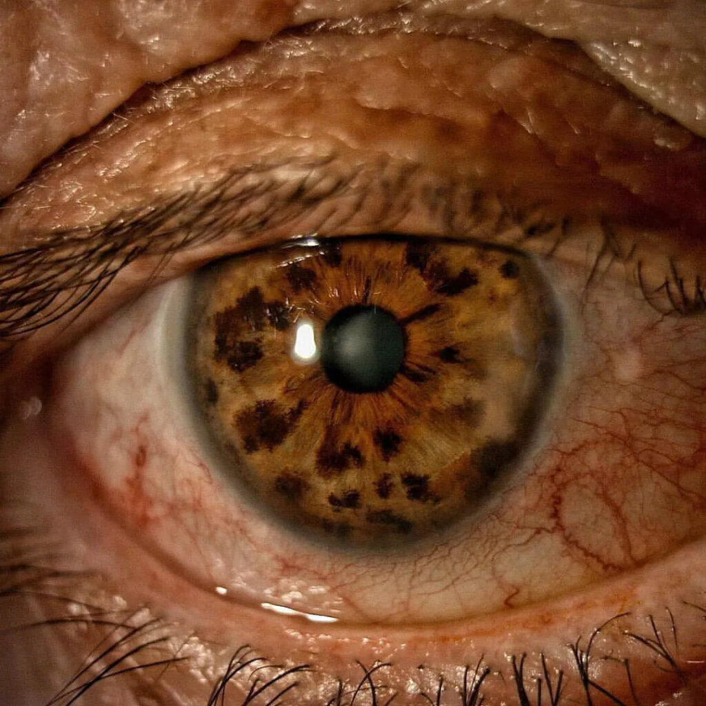
Iris freckles are small, flat brown spots on the anterior iris surface.
They are common, and most patients notice them only when someone points them out.
However, the clinical task is to confirm the lesion is a freckle and exclude a melanocytic tumor.
Usually, a careful slit lamp examination settles this in one visit.
What Iris Freckles Are
An iris freckle (ephelis) reflects extra melanin within melanocytes of the anterior border layer.
The number of melanocytes stays normal.
As a result, the spot has no mass and no thickness.
In contrast, a nevus contains a proliferation of nevus cells within the stroma.
How Common Brown Spots on the Iris Are
Freckles appear in many adults, and they become more frequent with age.
They are also easier to see in blue, green and light hazel irides.
In dark brown eyes, however, background pigment often hides them.
All-fit Slit-Lamp Adapter
Record and share exactly what you see at the slit lamp. One adapter fits any slit lamp or surgical microscope — and any smartphone.
From Choroida — the team behind this siteUV Exposure and Iris Freckles
Several studies link iris freckles with cumulative sunlight exposure.
For example, freckles tend to cluster in the inferior iris, which the upper lid does not shade.
However, this association does not prove that sunlight causes iris melanoma.
Iris Freckles and Uveal Melanoma Risk
Harbour and colleagues reported more iris freckles in patients with posterior uveal melanoma.
This finding suggests shared risk factors, such as light iris color and sun sensitivity.
Still, no evidence shows that a freckle itself transforms into melanoma.
Therefore, multiple freckles may simply flag a patient who deserves a careful dilated fundus exam.
Slit Lamp Signs of a Freckle
First, start with diffuse illumination to map the spots.
Then, use a narrow oblique beam to judge elevation.
Typical features
- Small size, often around 1 mm or less
- Flat surface with no measurable thickness
- Also, preserved crypts and normal stroma beneath the pigment
- Tan to dark brown color with soft edges
- Often multiple, scattered, and sometimes present in both eyes
Iris Freckles Versus Iris Nevus
A nevus is usually larger and more discrete, often a few millimeters across.
It may efface the crypts or slightly elevate the stroma.
Meanwhile, a freckle sits on the surface like ink on paper.
For a fuller comparison, review iris nevus versus melanoma.
Warning Signs of Melanoma
Iris melanoma is uncommon, but it is visible and often treatable when found early.
Shields and colleagues summarized the features predicting nevus growth into melanoma with the ABCDEF guide.
- Age young
- Blood (hyphema)
- Clock hour inferior
- Diffuse configuration
- Ectropion uveae
- Feathery tumor margins
In addition, intrinsic vessels, seeding, raised pressure and documented growth need prompt assessment (see iris melanoma).
Other Brown Spots to Consider
- Iris nevus, larger and more discrete
- For example, Lisch nodules in neurofibromatosis type 1 are dome-shaped and usually bilateral
- Iris mammillations, a uniform velvety surface pattern
- Also, ocular melanocytosis with diffuse hyperpigmentation
- Siderosis from a retained iron foreign body
Gonioscopy and Imaging
A typical freckle needs no extra testing.
However, perform gonioscopy when pigment approaches the angle or the pressure rises.
Ultrasound biomicroscopy then measures thickness and shows ciliary body extension.
When to Photograph Iris Freckles
Baseline slit lamp photography helps when freckles are numerous or the patient is anxious.
Also, use the same magnification and illumination at every visit (see smartphone slit lamp photography).
As a result, later comparisons rely on images instead of memory.
Follow-Up for Iris Freckles
A classic freckle needs no specific surveillance schedule.
However, record the location and number of spots in the chart.
Any change in size, shape or color then prompts comparison with baseline photos.
When to Refer
- Lesion larger than a typical freckle, or with measurable thickness
- Also, documented growth on serial photographs
- Feathery margins, intrinsic vessels or seeding
- In addition, hyphema, ectropion uveae or raised intraocular pressure
Counseling Patients About Iris Freckles
Most patients ask one direct question: harmless or not?
A flat freckle is a benign pigment change, and you can say so clearly.
Finally, ask patients to report any visible change in the spot.
Treatment of Iris Freckles
No treatment is needed.
Freckles do not affect vision, pupil function or eye pressure.
However, cosmetic procedures that alter iris color can cause inflammation and glaucoma.
Pitfalls to Avoid
- First, do not label a thick lesion a freckle without a slit-beam check
- Also, never skip the dilated fundus exam in patients with many freckles
- Then, avoid judging change from memory; photograph instead


Document what you see
Two smartphone imaging tools built for everyday clinic use — one for the slit lamp, one for the fundus.
From Choroida — the team behind this siteReferences
- Harbour JW, Brantley MA Jr, Hollingsworth H, Gordon M. Association between posterior uveal melanoma and iris freckles, iris naevi, and choroidal naevi. Br J Ophthalmol. 2004;88:36-38.
- Shields CL, Kaliki S, Hutchinson A, et al. Iris nevus growth into melanoma: analysis of 1611 consecutive eyes: the ABCDEF guide. Ophthalmology. 2013;120:766-772.
- Shields CL, Kaliki S, Shah SU, et al. Iris melanoma: features and prognosis in 317 children and adults. J AAPOS. 2012;16:10-16.
- American Academy of Ophthalmology. Basic and Clinical Science Course, Section 4: Ophthalmic Pathology and Intraocular Tumors. San Francisco: AAO; latest edition.
- Bowling B. Kanski’s Clinical Ophthalmology: A Systematic Approach. 8th ed. Elsevier; 2016.
Test yourself
A few questions straight from this article.
-
What is the histologic basis of an iris freckle (ephelis)?
An iris freckle reflects extra melanin within melanocytes of the anterior border layer while their number stays normal, so the spot has no mass or thickness. -
Which histologic feature characterizes an iris nevus rather than an iris freckle?
A nevus contains a proliferation of nevus cells within the stroma, whereas a freckle has a normal melanocyte count with extra pigment. -
What is the typical size of an iris freckle?
Freckles are small, often around 1 mm or less, while a nevus is usually larger and more discrete, often a few millimeters across. -
Which slit-lamp technique best judges elevation of a suspected iris freckle?
Diffuse illumination maps the spots, then a narrow oblique beam judges elevation; a true freckle is flat with preserved crypts and normal stroma beneath the pigment. -
Which examination is warranted in a patient with multiple iris freckles, given their reported association with posterior uveal melanoma?
More iris freckles have been reported in patients with posterior uveal melanoma, suggesting shared risk factors, though no evidence shows a freckle transforms; multiple freckles may simply flag the need for a careful dilated fundus exam. -
In the ABCDEF guide to iris nevus growth into melanoma, what does the letter F denote?
The guide lists young age, blood (hyphema), inferior clock hour, diffuse configuration, ectropion uveae and feathery tumor margins as features predicting nevus growth into melanoma. -
Which brown iris lesion of neurofibromatosis type 1 can mimic iris freckles but is dome-shaped and usually bilateral?
Lisch nodules in neurofibromatosis type 1 are dome-shaped and usually bilateral, whereas iris mammillations form a uniform velvety surface pattern and a freckle stays flat. -
Which test measures thickness and shows ciliary body extension when an iris pigmented lesion raises concern?
A typical freckle needs no extra testing, but when pigment approaches the angle or pressure rises, gonioscopy is performed and ultrasound biomicroscopy measures thickness and ciliary body extension. -
What follow-up does a classic iris freckle need?
A classic freckle needs no specific surveillance schedule, but its location and number are recorded so that any change prompts comparison with baseline photographs. -
Which finding means a presumed iris freckle should be referred?
Referral is indicated for a lesion larger than a typical freckle or with measurable thickness, documented growth, feathery margins, intrinsic vessels, seeding, hyphema, ectropion uveae or raised pressure.

Writes and reviews the clinical articles on Educate. Every post is checked against peer-reviewed sources before it goes live.