OCT retinal layers appear as alternating reflective and dark bands that correspond to cell bodies, synapses and fibers.
Reading them well requires knowing the anatomy and the names agreed by consensus, so this article lists the layers from the vitreous to the choroid.
Why Learn OCT Retinal Layers
Every macular disease changes one or more layers, so naming the layer helps you describe the lesion and its prognosis.
A common language also allows clear communication, and because the consensus names changed over time, you should use the current terms.
The Inner Retina Among OCT Retinal Layers
The retinal nerve fiber layer lies next to the vitreous and is bright, and the ganglion cell layer and the inner plexiform layer follow.
The inner nuclear layer is dark because it contains cell bodies, so the inner retina shows a pattern of bright and dark bands.
Fundus Explorer Pro
Photograph the retinal findings described here with the phone already in your pocket — 22 D optics and built-in illumination in one handheld unit.
From Choroida — the team behind this siteOuter Plexiform Layer and Henle Fiber Layer
The outer plexiform layer lies between the inner and outer nuclear layers, and at the fovea the Henle fiber layer lies in the same space and runs obliquely.
Lujan and colleagues showed that its reflectivity depends on the angle of the beam, which explains why the layer looks different away from the center.
The Outer Nuclear Layer
The outer nuclear layer holds the nuclei of the photoreceptors and is dark and thick at the fovea. Thinning of this layer means loss of photoreceptors, so it is a marker of outer retinal damage.
The Four Outer Retinal Bands
The IN-OCT consensus named four bands in the outer retina, which are the external limiting membrane, the ellipsoid zone, the interdigitation zone and the retinal pigment epithelium with Bruch membrane. Staurenghi and colleagues proposed the names in 2014, and they replaced the older terms for the inner segment and outer segment junction.
The External Limiting Membrane
The external limiting membrane is the first band of the outer retina, a thin line formed by junctions between Muller cells and photoreceptors. Its loss predicts poor visual recovery in several conditions, and because it is subtle, you should look for it on high-quality scans.
The Ellipsoid Zone
The second band represents the mitochondria-rich ellipsoid of the inner segments, an assignment Spaide and Curcio proposed in 2011. Jonnal and colleagues also examined the cellular origin of the bands in adaptive optics, and therefore disruption of this band is a marker of photoreceptor damage (see ellipsoid zone disruption).
Interdigitation Zone and RPE
The third band reflects the contact between cone outer segments and RPE processes, while the fourth band reflects the RPE and Bruch membrane. A gap between them may appear in disease, and because the third band is faint, it needs good image quality.
The Foveal Pit and Bouquet
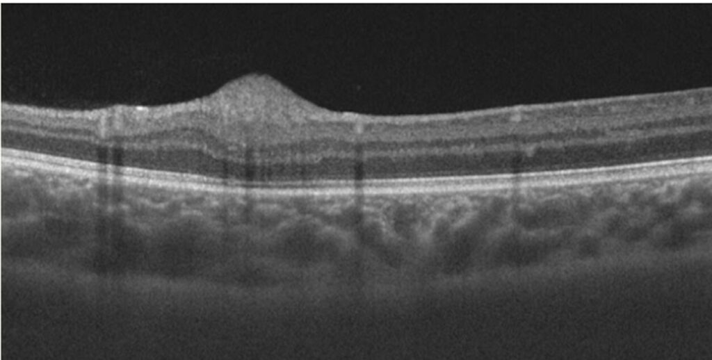
The fovea has a pit where inner layers are displaced, and the bouquet of cone outer segments gives a characteristic shape (see central foveal bouquet). The pit also varies with the individual, so interpret a foveal shape in context.
Choroid in OCT Retinal Layers
The choroid lies beneath the RPE and appears dark with vessel lumens. Enhanced depth imaging and swept-source devices show it well, and the sclerochoroidal junction marks the outer border.
Segmentation Errors in OCT Retinal Layers
Software draws boundaries and can fail in disease, so check the lines on the B-scan when thickness values look odd. Manual correction also helps in research and in complex cases, and because errors are common, do not rely on the numbers alone.
Using OCT Retinal Layers in Diagnosis
Match the abnormal layer with the disease. Outer retinal disruption suggests photoreceptor disease or trauma (see outer retinal tubulation), while inner layer changes suggest ischemia or glaucoma. Therefore, a layer-based approach organizes the differential.
Scanning Tips
Center the scan on the fovea and check the focus, and use a raster or radial protocol for the macula. Average repeated scans to reduce noise, and because image quality limits layer detail, repeat poor scans.
Reflectivity and Terminology
Hyperreflective means brighter than the surrounding layer, while hyporeflective means darker. Shadows from vessels and opacities can also mimic loss, so learn to recognize shadowing.
Layers in Disease Examples
In macular hole, the layers separate and retract, and in macular telangiectasia, cavities appear in the outer retina. In geographic atrophy, the outer layers vanish, so describe the findings by layer.


Document what you see
Two smartphone imaging tools built for everyday clinic use — one for the slit lamp, one for the fundus.
From Choroida — the team behind this siteReferences
- Staurenghi G, Sadda S, Chakravarthy U, Spaide RF. Proposed lexicon for anatomic landmarks in normal posterior segment spectral-domain optical coherence tomography: the IN-OCT consensus. Ophthalmology. 2014;121:1572-1578.
- Spaide RF, Curcio CA. Anatomical correlates to the bands seen in the outer retina by optical coherence tomography: literature review and model. Retina. 2011;31:1609-1619.
- Jonnal RS, Kocaoglu OP, Zawadzki RJ, Lee SH, Werner JS, Miller DT. The cellular origins of the outer retinal bands in optical coherence tomography images. Invest Ophthalmol Vis Sci. 2014;55:7904-7918.
- Lujan BJ, Roorda A, Knighton RW, Carroll J. Revealing Henle’s fiber layer using spectral domain optical coherence tomography. Invest Ophthalmol Vis Sci. 2011;52:1486-1492.
Test yourself
A few questions straight from this article.
-
On a normal macular OCT scan, which bright layer lies immediately next to the vitreous?
Reading OCT layers from the vitreous inward, the bright retinal nerve fiber layer comes first, followed by the ganglion cell and inner plexiform layers. -
On OCT, what makes the reflectivity of the Henle fiber layer vary across the macula?
The Henle fibers run obliquely, and their reflectivity depends on the beam angle, which explains why the layer looks different away from the foveal center. -
Thinning of the outer nuclear layer on OCT indicates loss of which cells?
The outer nuclear layer holds the photoreceptor nuclei and is dark and thick at the fovea, so its thinning marks photoreceptor loss and outer retinal damage. -
Under the IN-OCT consensus, which older OCT term did the ellipsoid zone replace?
The IN-OCT consensus named four outer retinal bands, and its terms replaced the older label of the inner segment and outer segment junction. -
The second outer retinal band on OCT, the ellipsoid zone, corresponds to which photoreceptor structure?
The ellipsoid zone represents the mitochondria-rich ellipsoid of the photoreceptor inner segments, and disruption of this band marks photoreceptor damage. -
The external limiting membrane on OCT is formed by junctions between which cells?
The external limiting membrane is a thin line formed by Muller cell–photoreceptor junctions; it is subtle, and its loss predicts poor visual recovery in several conditions. -
The third outer retinal band on OCT, the interdigitation zone, reflects contact between which structures?
The interdigitation zone reflects cone outer segments meeting RPE processes; it is faint and needs good image quality, while the fourth band is the RPE–Bruch membrane complex. -
On OCT imaging of the choroid, which landmark marks its outer border?
The choroid lies beneath the RPE and appears dark with vessel lumens; enhanced depth imaging and swept-source OCT show it well, with the sclerochoroidal junction as its outer border. -
When automated OCT retinal layer thickness values look implausible, what should be checked first?
Segmentation software can fail in disease, so check the drawn lines on the B-scan and correct them manually when needed rather than relying on the numbers alone. -
On OCT, abnormalities confined to the inner retinal layers most suggest which category of disease?
A layer-based approach organizes the differential: inner layer changes point to ischemia or glaucoma, whereas outer retinal disruption suggests photoreceptor disease or trauma.

Writes and reviews the clinical articles on Educate. Every post is checked against peer-reviewed sources before it goes live.