Metamorphopsia is the perception that straight lines look bent, wavy, or broken, and it usually signals macular disease.
Patients often describe it as a distorted face, a bent door frame, or letters that shift on the page. The symptom persists in many patients after successful treatment, so clinicians should measure it along with visual acuity.
What Metamorphopsia Means
Metamorphopsia arises when photoreceptors move closer together, farther apart, or out of their normal alignment. The brain maps the signals from displaced receptors to their old positions, so the image bends. Vertical and horizontal lines may distort differently, which helps the clinician to understand the direction of traction or fluid.
Causes of Metamorphopsia
- Epiretinal membrane and vitreomacular traction
- Macular edema and central serous disease
- Neovascular age-related macular degeneration
- Macular hole and lamellar hole
Less common causes include retinal detachment involving the macula, choroidal tumors, and high myopia with macular schisis (see epiretinal membrane).
Fundus Explorer Pro
Photograph the retinal findings described here with the phone already in your pocket — 22 D optics and built-in illumination in one handheld unit.
From Choroida — the team behind this siteTesting Metamorphopsia
The Amsler grid is the usual first test, though its sensitivity is limited. M-CHARTS use a series of dotted lines with increasing dot spacing, and they give a numerical score.
Matsumoto and colleagues showed that M-CHARTS quantify the distortion in epiretinal membrane, and Arimura and colleagues applied them to macular holes. Wiecek and colleagues later developed other quantitative tests that map the shape of the distortion (see full-thickness macular hole surgery).
Metamorphopsia After Treatment
Distortion often improves more slowly than acuity. Many patients keep some degree of metamorphopsia after membrane peeling or hole closure, and the outer retinal layers may explain this.
Eyes with less preoperative distortion and a shorter history tend to recover better. Therefore, early surgery may help to protect against lasting distortion (see why some macular holes reopen).
Managing Metamorphopsia
Treat the underlying cause, such as a membrane, fluid, or neovascularization, and explain that vision may continue to improve for six to twelve months. Offer reassurance, and use simple advice such as viewing grids with one eye at a time. Refer patients with persistent distortion for low vision support when function is limited.
Differential Diagnosis of Metamorphopsia
Optic nerve disease does not usually cause line distortion, so look for macular findings. Corneal irregularity, cataract, and refractive error can mimic the symptom, so check refraction and use a pinhole, and OCT separates most causes within minutes.
An Example Case
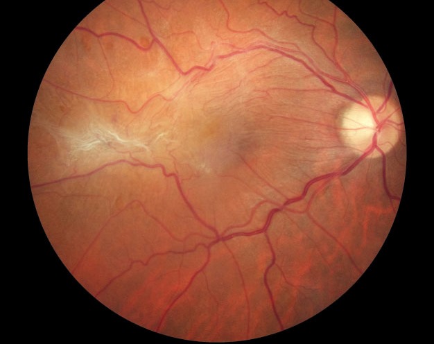
A 67-year-old woman reports that the straight edge of her kitchen cabinets looks wavy in her right eye. Her acuity is 20/30, and OCT shows an epiretinal membrane with thickening and a loss of the foveal contour.
She scores 0.8 on the M-CHARTS vertical line test, and she notes trouble reading. After vitrectomy and membrane peeling, her acuity reaches 20/25, and the score falls to 0.4 over a year. She still notices a mild bend, but the distortion no longer disturbs her reading.
Questions Patients Ask
Patients ask whether the distortion will disappear completely, and the honest answer is that it improves in most eyes but may not vanish. They also ask whether the other eye will develop the same problem, and it can, since membranes are often bilateral. Because some patients feel anxious, explain that the symptom itself does not harm the retina.
Pitfalls to Avoid
- Relying on acuity alone to judge outcome after macular surgery
- Missing early membranes because the Amsler grid looked normal
- Attributing distortion to the cornea without checking the macula
- Promising full recovery of distortion before surgery
Looking Ahead
Researchers study OCT features, such as inner retinal wrinkling, that predict the degree of distortion. Also, tablet-based tests may allow home monitoring of distortion, and therefore patients may soon track their own recovery between visits.
Practical Points
Ask patients to look at a door frame or a grid with each eye separately. Then measure with a quantitative chart when you plan surgery, and repeat it afterward. Also record the date of onset, since longer duration predicts slower recovery.
How Distortion Changes Over Time
Distortion often peaks before surgery and then falls gradually, and because inner retinal layers may remodel, improvement can continue for a year or more. Also, patients with bilateral disease may notice a second eye later, so review both eyes at each visit and use the same test each time.
Self-Monitoring
Teach patients with early membranes to check a grid weekly with each eye. If lines become more bent, ask them to call the clinic. Because change often precedes acuity loss, early reports help to time surgery.


Document what you see
Two smartphone imaging tools built for everyday clinic use — one for the slit lamp, one for the fundus.
From Choroida — the team behind this siteReferences
- Matsumoto C, Arimura E, Okuyama S, et al. Quantification of metamorphopsia in patients with epiretinal membranes. Invest Ophthalmol Vis Sci. 2003;44:4012-4016.
- Arimura E, Matsumoto C, Okuyama S, et al. Quantification of metamorphopsia in a macular hole patient using M-CHARTS. Acta Ophthalmol Scand. 2007;85:55-59.
- Wiecek E, Lashkari K, Dakin SC, Bex P. Novel quantitative assessment of metamorphopsia in maculopathy. Invest Ophthalmol Vis Sci. 2015;56:494-504.
- American Academy of Ophthalmology. Basic and Clinical Science Course, Section 12: Retina and Vitreous. AAO; current edition.
Test yourself
A few questions straight from this article.
-
Metamorphopsia, the perception that straight lines look bent or wavy, usually signals disease of which structure?
Metamorphopsia usually signals macular disease. Optic nerve disease does not usually cause line distortion, so the clinician should look for macular findings. -
What mechanism produces metamorphopsia in macular disease?
When photoreceptors move closer together, farther apart or out of alignment, the brain still maps their signals to the original positions, so the image bends. -
In metamorphopsia, what can it tell the clinician when vertical and horizontal lines distort differently?
Vertical and horizontal lines may distort differently, and the difference helps the clinician understand which way the traction or fluid is acting. -
What limitation of the Amsler grid matters when screening for metamorphopsia from an early epiretinal membrane?
The Amsler grid is the usual first test, but its sensitivity is limited. Missing early membranes because the grid looked normal is a recognised pitfall. -
How do M-CHARTS measure metamorphopsia?
M-CHARTS present a series of dotted lines with increasing dot spacing and give a numerical score. They have been used to quantify distortion in epiretinal membrane and macular hole. -
After epiretinal membrane peeling or macular hole closure, how does metamorphopsia typically recover compared with visual acuity?
Distortion often improves more slowly than acuity, and many patients keep some metamorphopsia after surgery. That is why acuity alone should not be used to judge the outcome. -
Which preoperative features predict better recovery from metamorphopsia after macular surgery?
Eyes with less preoperative distortion and a shorter history tend to recover better. On that basis, early surgery may help protect against lasting distortion. -
Corneal irregularity, cataract and refractive error can mimic metamorphopsia. Which simple steps help to exclude them?
Anterior segment and refractive causes can mimic line distortion, so check the refraction and use a pinhole. OCT then separates most macular causes within minutes. -
Why are patients with early epiretinal membranes taught to check a grid weekly, one eye at a time?
Patients should call the clinic if the lines become more bent. Change in distortion often comes before acuity loss, so early reports help with timing surgery. -
In a patient with metamorphopsia from an epiretinal membrane, why should both eyes be reviewed at each visit?
Membranes are often bilateral, and patients with bilateral disease may notice the second eye later. Review both eyes at each visit and use the same test each time.

Writes and reviews the clinical articles on Educate. Every post is checked against peer-reviewed sources before it goes live.