Swept-source OCT uses a tunable laser at about 1050 nm, and it images deeper tissue and a wider field than spectral-domain systems.
The longer wavelength penetrates the retinal pigment epithelium, cataract, and hemorrhage better, so these features make it useful for choroidal disease, high myopia, and wide-field imaging.
How Swept-Source OCT Works
The laser source sweeps rapidly through a range of wavelengths and a single photodetector records the interference signal, while spectral-domain systems use a broadband light source and a spectrometer instead.
Swept-source devices scan at 100,000 A-scans per second or faster, which reduces motion artifacts and allows wide scans, and longer wavelengths also scatter less in tissue, so sensitivity roll-off with depth is lower (see OCT angiography).
Choroidal Imaging With Swept-Source OCT
The choroid appears more clearly because less signal is lost in the pigment epithelium, and Povazay and colleagues showed the benefits of longer wavelengths for choroidal vessels in early work.
Studies of choroidal thickness give similar results with swept-source and enhanced depth spectral-domain methods, with some differences in the measured values. Clinicians use the images in pachychoroid disease, polypoidal vasculopathy, and central serous disease (see pachychoroid neovasculopathy).
Fundus Explorer Pro
Photograph the retinal findings described here with the phone already in your pocket — 22 D optics and built-in illumination in one handheld unit.
From Choroida — the team behind this siteWide-Field Swept-Source OCT
Modern devices scan fields of 12 by 12 mm or larger in a single image, and wide scans show the macula, the optic disc, and the posterior pole together. Surgeons and clinicians use them to study staphylomas, optic disc pits, and peripapillary changes (see ultra-widefield retinal imaging).
Swept-Source OCT Angiography
Fast scanning gives dense angiographic data with fewer motion artifacts, and wide-angle OCT angiography reveals nonperfusion and neovascularization across larger fields. Therefore, many centers adopt swept-source platforms for angiography.
Swept-Source OCT Versus Spectral-Domain OCT
- Swept-source images deeper layers and sees through media opacities better
- Spectral-domain gives higher axial resolution in some devices
- Swept-source costs more and offers fewer normative databases
- Both give good results for the macula in most patients
For routine macular disease, either type serves well.
Limits
Segmentation software may misplace boundaries in the choroid, so check images manually. Norms differ between devices, so avoid mixing measurements from different instruments, and artifacts still occur, especially with media opacities and eye movement.
An Example Case
A 68-year-old man has blurred vision from a dense cataract and a suspected macular lesion, and spectral-domain OCT gives a poor image because of the media opacity.
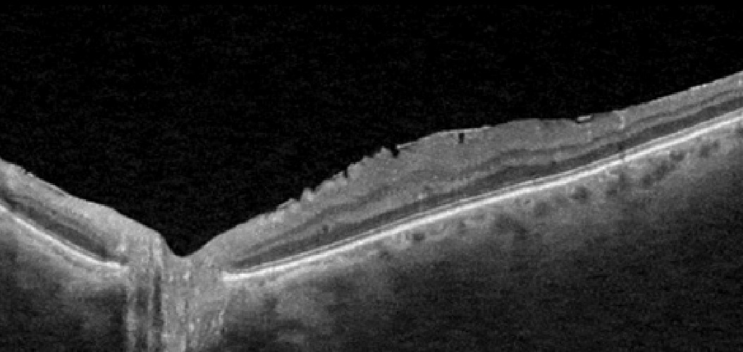
Swept-source OCT penetrates the cataract and shows a polypoidal lesion with a branching vascular network under the RPE, and the findings guide indocyanine green angiography and treatment planning.
Questions Clinicians Ask
Clinicians ask whether swept-source OCT replaces spectral-domain devices, and many clinics use both. They also ask about the cost, and swept-source systems usually cost more, though prices continue to fall. Because most macular disease images well with either type, choose by case mix.
Pitfalls to Avoid
- Comparing thickness values between different devices without conversion
- Trusting automated choroidal segmentation without checking the boundaries
- Ignoring signal strength in dense media
- Assuming deeper penetration always gives clearer detail
Looking Ahead
Faster lasers will increase scan area and detail further, and integration with artificial intelligence may speed up interpretation. However, normative databases need time to catch up with the new instruments.
Practical Points
Use the same device for follow-up in a given patient, and document the scan pattern and size in the notes. Also use wide scans when the question involves the disc, the staphyloma, or the peripapillary choroid.
Choosing Scan Patterns
Select a wide scan to see the posterior pole in one image, and a radial or raster pattern to study the macula in detail. Then use angiography scans of 6 by 6 or 12 by 12 mm according to the question. Also use enhanced depth modes only when needed, since they lower image quality in the retina.
Reading Choroidal Images
Measure thickness from the RPE to the sclerochoroidal interface, and note any dilated vessels, but verify the boundary manually because it may be unclear. Pachychoroid disease shows a thick choroid with dilated outer vessels and thin inner layers. Therefore, a careful look at the choroid supports diagnosis and treatment choices.
Document the scan size in the report, since it changes what the image can show. Also teach the team to label the scan type in each report, so that future comparisons use the same settings, and plan storage and backup because wide scans create large files.


Document what you see
Two smartphone imaging tools built for everyday clinic use — one for the slit lamp, one for the fundus.
From Choroida — the team behind this siteReferences
- Povazay B, Bizheva K, Hermann B, et al. Enhanced visualization of choroidal vessels using ultrahigh resolution ophthalmic OCT at 1050 nm. Opt Express. 2003;11:1980-1986.
- Copete S, Flores-Moreno I, Montero JA, et al. Direct comparison of spectral-domain and swept-source OCT in the measurement of choroidal thickness in normal eyes. Br J Ophthalmol. 2014;98:334-338.
- Spaide RF, Fujimoto JG, Waheed NK, et al. Optical coherence tomography angiography. Prog Retin Eye Res. 2018;64:1-55.
- American Academy of Ophthalmology. Basic and Clinical Science Course, Section 12: Retina and Vitreous. AAO; current edition.
Test yourself
A few questions straight from this article.
-
At approximately what wavelength does the tunable laser of swept-source OCT operate?
Swept-source OCT uses a tunable laser at about 1050 nm, a longer wavelength that lets it image deeper tissue and a wider field than spectral-domain systems. -
In swept-source OCT, which component records the interference signal as the laser sweeps through its wavelengths?
The swept laser runs rapidly through a range of wavelengths while a single photodetector records the interference; spectral-domain systems instead pair a broadband source with a spectrometer. -
What scanning speed do swept-source OCT devices typically reach?
Swept-source devices scan at 100,000 A-scans per second or faster, which reduces motion artifacts and makes wide scans possible. -
Why does swept-source OCT show the choroid more clearly than spectral-domain OCT?
The longer wavelength penetrates the retinal pigment epithelium better, so less signal is lost there and the choroidal vessels appear more clearly. -
Compared with swept-source OCT, what advantage can some spectral-domain OCT devices offer?
Spectral-domain OCT gives higher axial resolution in some devices, whereas swept-source images deeper layers and sees through media opacities better. -
What field size can modern wide-field swept-source OCT devices capture in a single scan?
Modern swept-source devices scan fields of 12 by 12 mm or larger in one image, showing the macula, optic disc and posterior pole together. -
Between which boundaries is choroidal thickness measured on swept-source OCT?
Choroidal thickness is measured from the RPE to the sclerochoroidal interface, and that boundary should be verified manually because it may be unclear. -
How should automated choroidal segmentation on swept-source OCT be handled?
Segmentation software may misplace boundaries in the choroid, so the images should be checked manually rather than trusted blindly. -
When tracking choroidal measurements over time in one patient, what practice is recommended for swept-source OCT?
Norms differ between devices, so measurements from different instruments should not be mixed and the same device should be used for a given patient's follow-up. -
What swept-source OCT choroidal appearance characterises pachychoroid disease?
Pachychoroid disease shows a thick choroid with dilated outer vessels and thinned inner choroidal layers, which careful choroidal reading picks up.

Writes and reviews the clinical articles on Educate. Every post is checked against peer-reviewed sources before it goes live.