Conjunctival melanoma is a malignant tumor of the melanocytes of the conjunctiva, and it accounts for a small share of ocular melanomas.
It arises most often from primary acquired melanosis with atypia, less often from a nevus, and sometimes de novo.
Early recognition and complete removal are the main factors that improve the outcome, because recurrence and spread are real risks.
What Is Conjunctival Melanoma?
The tumor typically affects middle-aged and older adults, and it is more frequent in fair-skinned people.
It can also arise on the bulbar conjunctiva, the limbus, the fornix, the palpebral conjunctiva, or the caruncle.
It may extend to the cornea or the eyelid.
In contrast with uveal melanoma, it behaves more like cutaneous melanoma, and researchers have found mutations in BRAF and NRAS in a proportion of tumors.
Doctors suspect ultraviolet exposure as a risk factor, especially for tumors arising on the exposed conjunctiva.
The tumor spreads to regional lymph nodes first, and then to distant sites.
Clinical Features of Conjunctival Melanoma
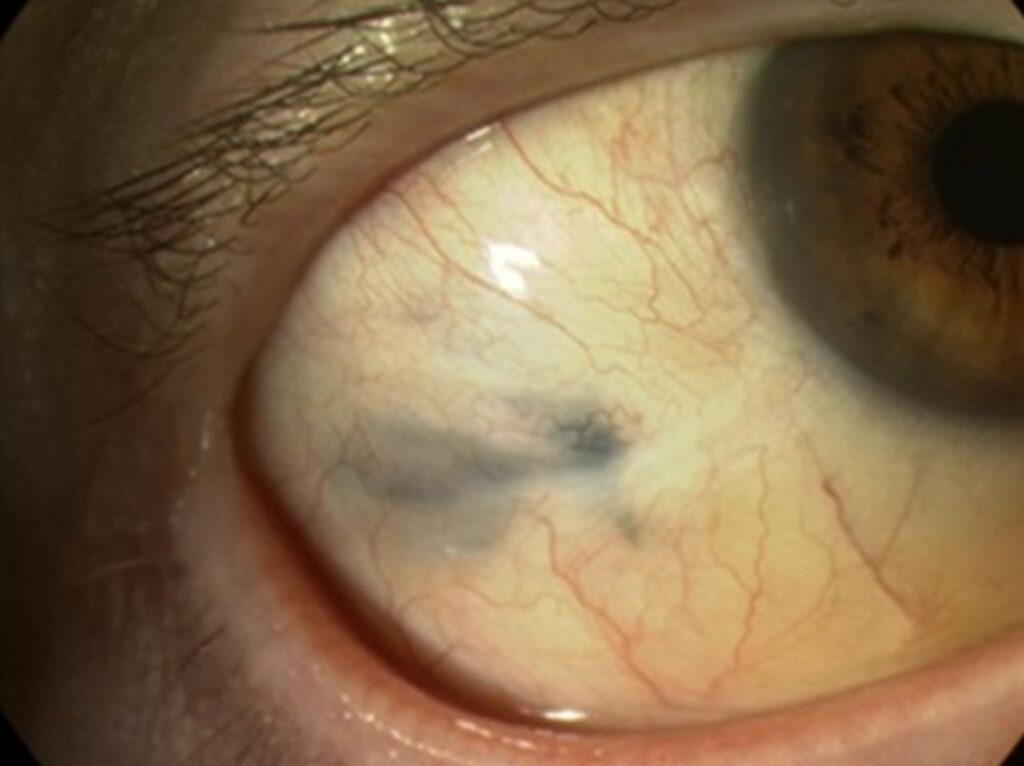
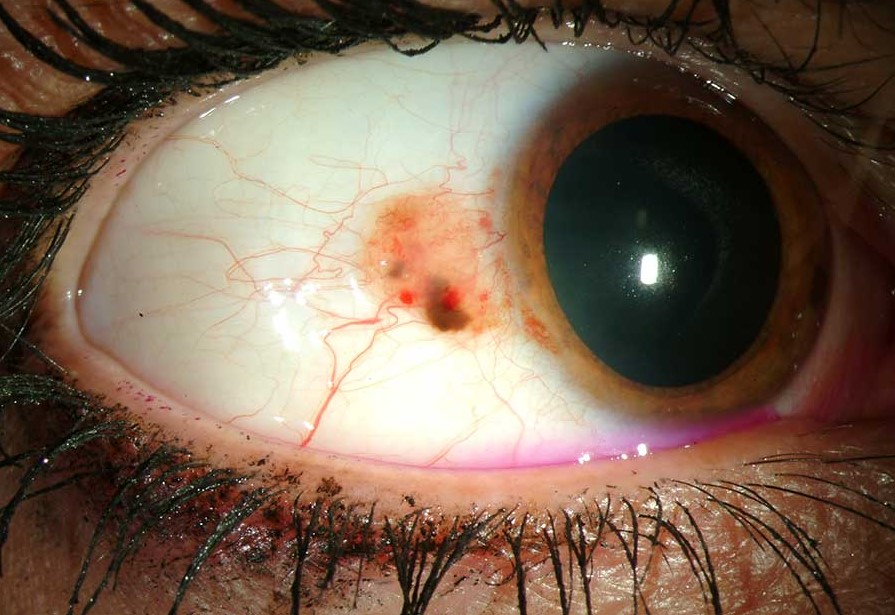
- A pigmented, nodular, or fleshy lesion, which may be brown, black, or pink and amelanotic
- Prominent feeder vessels and surface vascularity
- Irregular or ill-defined edges, sometimes with adjacent flat pigmentation
- Growth over months, change in color, or bleeding
- Location at the limbus or in the fornix, where it may hide
- Associated primary acquired melanosis in many cases (see conjunctival nevus)
Evert the lids and examine the fornices and the caruncle in every patient with a pigmented lesion, because tumors can hide under the lids.
All-fit Slit-Lamp Adapter
Record and share exactly what you see at the slit lamp. One adapter fits any slit lamp or surgical microscope — and any smartphone.
From Choroida — the team behind this siteDifferential Diagnosis of Conjunctival Melanoma
- Conjunctival nevus: well-defined, often cystic, stable over years
- Primary acquired melanosis: flat, patchy, and ill-defined pigment
- Racial melanosis and secondary melanosis: from inflammation or drugs
- Pigmented squamous neoplasia: a lesion that can look dark (see conjunctival intraepithelial neoplasia)
- Scleral and episcleral pigment: deeper and blue-gray, not mobile with the conjunctiva
- Foreign body or hemorrhage: which can mimic pigment
Evaluation for Conjunctival Melanoma
Document the lesion with photographs and measurements, and map its extent.
Also, examine the regional lymph nodes in the preauricular and cervical areas.
Anterior segment OCT and ultrasound biomicroscopy can show the thickness and the depth of invasion.
Take a biopsy if the diagnosis is uncertain, but plan it so that you do not spread the tumor.
Consider staging with imaging for thicker tumors, and surgeons may use sentinel lymph node biopsy in selected cases.
Treatment
The primary treatment is wide excision using a no-touch technique, which avoids direct contact with the tumor and uses a margin of normal conjunctiva.
Cryotherapy to the conjunctival margins, alcohol application to the cornea, and sometimes the use of a plaque or brachytherapy reduce the chance of local recurrence.
Topical chemotherapy with mitomycin C or interferon can treat residual disease or associated acquired melanosis.
Larger tumors with orbital involvement may require exenteration, though this is now less common.
Doctors use newer systemic therapies, such as checkpoint inhibitors and targeted agents, for advanced disease, on the basis of experience with cutaneous melanoma.
Prognosis and Follow-Up
Thickness is the most important prognostic factor, and tumors thicker than a few millimeters have a higher risk of metastasis.
In addition, other adverse features include nonbulbar location, caruncular involvement, and incomplete excision.
Patients need lifelong follow-up, with examination of the ocular surface and the lymph nodes at regular intervals, and referral to an oncologist when indicated.
The No-Touch Technique
The surgeon avoids direct contact with the tumor and handles tissue only at the margins, using a margin of several millimeters of clear conjunctiva.
The surgeon lifts the tumor in a single piece with the underlying Tenon tissue when necessary, and changes instruments that the tumor contaminated.
Also, the surgeon treats the conjunctival edges with cryotherapy using a double freeze-thaw cycle.
Clinicians may treat the corneal epithelium with absolute alcohol.
Surgeons reconstruct with amniotic membrane or conjunctival grafts, taking from the same eye only after confirming tumor-free margins (see amniotic membrane transplantation).
Pathology and Staging
Orient the specimen and send it with a clinical history, so that the pathologist can assess margins and thickness.
Also, the AJCC staging system for conjunctival melanoma uses tumor thickness, location, and involvement of nearby structures.
Sentinel node biopsy can detect microscopic nodal disease, though its role is debated and depends on local expertise.
Follow-Up and Systemic Care
Review patients every three to six months for the first years, with examination of the conjunctival surface under magnification and palpation of the nodes.
The stage guides periodic imaging, and the care of advanced disease involves an ocular oncologist and a medical oncologist.
Clinicians often see recurrences at the surgical site or the fornix, and early detection allows less extensive treatment.
Educate the patient to report new pigmentation or growth, since lesions can recur in different parts of the conjunctiva.
For the latest studies, see the PubMed results for conjunctival melanoma.


Document what you see
Two smartphone imaging tools built for everyday clinic use — one for the slit lamp, one for the fundus.
From Choroida — the team behind this siteReferences
- Shields CL, Markowitz JS, Belinsky I, et al. Conjunctival melanoma: outcomes based on tumor origin in 382 consecutive cases. Ophthalmology. 2011;118:389-395.
- Shields CL, Shields JA, Gündüz K, et al. Conjunctival melanoma: risk factors for recurrence, exenteration, metastasis, and death in 150 consecutive patients. Trans Am Ophthalmol Soc. 2000;98:471-483.
- Shields JA, Shields CL. Eyelid, Conjunctival, and Orbital Tumors: An Atlas and Textbook. 3rd ed. Wolters Kluwer; 2016.
- American Academy of Ophthalmology. Basic and Clinical Science Course, Section 4: Ophthalmic Pathology and Intraocular Tumors. AAO; current edition.
Test yourself
A few questions straight from this article.
-
From which precursor does conjunctival melanoma most often arise?
Conjunctival melanoma arises most often from primary acquired melanosis with atypia, less often from a nevus, and sometimes de novo. -
Unlike uveal melanoma, conjunctival melanoma behaves more like which tumour?
Conjunctival melanoma behaves more like cutaneous melanoma, with BRAF and NRAS mutations found in a proportion of tumours. -
Where does conjunctival melanoma spread first?
The tumour spreads first to regional lymph nodes and then to distant sites, so preauricular and cervical nodes are examined. -
In a patient with a pigmented conjunctival lesion, why should the lids be everted and the fornices examined for conjunctival melanoma?
Melanoma may sit in the fornix, palpebral conjunctiva or caruncle, so lids are everted and fornices and caruncle examined in every pigmented lesion. -
Which features favour a conjunctival nevus over conjunctival melanoma?
A nevus is well defined, often cystic and stable over years, whereas melanoma may show feeder vessels, ill-defined edges, growth or bleeding. -
How does scleral or episcleral pigment differ from conjunctival melanoma on examination?
Scleral and episcleral pigment lies deeper, looks blue-gray and does not move with the conjunctiva. -
What is the primary treatment for conjunctival melanoma?
Primary treatment is wide no-touch excision, avoiding direct tumour contact and taking a margin of several millimetres of clear conjunctiva. -
After no-touch excision of conjunctival melanoma, how are the conjunctival margins treated to reduce local recurrence?
Conjunctival edges are treated with double freeze-thaw cryotherapy, and the corneal epithelium may be treated with absolute alcohol. -
What is the most important prognostic factor in conjunctival melanoma?
Thickness is the most important prognostic factor; thicker tumours carry a higher risk of metastasis. -
Which feature is an adverse prognostic factor in conjunctival melanoma?
Besides thickness, adverse features include nonbulbar location, caruncular involvement and incomplete excision.

Writes and reviews the clinical articles on Educate. Every post is checked against peer-reviewed sources before it goes live.