
A corneal foreign body is among the most common reasons for an urgent ophthalmic visit.
While most cases are straightforward to manage, the details of the history and exam matter enormously: the difference between a superficial metallic fragment from grinding and a high-velocity projectile from hammering metal-on-metal is the difference between a routine office removal and a globe that needs urgent imaging to rule out intraocular penetration.
History: The Mechanism Changes Everything
The single most important question is how the foreign body arrived at the eye: low-velocity mechanisms (wind-blown debris, gardening, most workplace dust or particles) carry minimal risk of intraocular penetration.
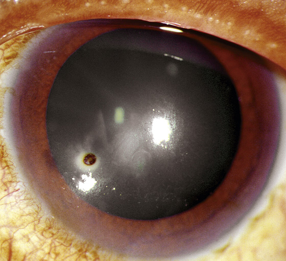
Meanwhile, high-velocity mechanisms — hammering metal on metal, grinding, drilling without eye protection — carry a genuine risk that a fragment has penetrated the globe and lodged intraocularly, sometimes with a deceptively small, easily overlooked entry wound.
Any history involving metal-on-metal contact at high speed should prompt a specific, deliberate search for signs of penetration before assuming the injury is limited to the corneal surface.
Asking specifically about the exact activity at the moment of injury — hammering, grinding, using a lawnmower or weed trimmer, or simply walking outdoors in windy conditions — is far more useful than a general question about how the eye was hurt, since patients do not always volunteer the mechanism details that actually determine the level of concern.
Clinical Presentation
Patients report the sudden onset of foreign-body sensation, tearing, redness, photophobia, and blepharospasm, usually with a clear description of when and how the injury occurred.
Vision may be reduced if the foreign body sits within the visual axis or if associated corneal edema and inflammation are affecting acuity, though vision is often relatively preserved with a small, peripherally located foreign body, making the location of the fragment relative to the visual axis a useful thing to note early in the exam.
All-fit Slit-Lamp Adapter
Record and share exactly what you see at the slit lamp. One adapter fits any slit lamp or surgical microscope — and any smartphone.
From Choroida — the team behind this siteExam Findings
- Visible foreign material embedded in the corneal surface, sometimes obvious, sometimes subtle and requiring careful slit-lamp examination with good illumination
- A rust ring, when the foreign body is iron- or steel-containing and has been present long enough for oxidation to begin, appearing as a brownish discoloration surrounding the entry site
- Surrounding corneal edema and an inflammatory infiltrate around the foreign body
- Fluorescein staining of the surrounding epithelial defect
- Careful examination for signs of full-thickness penetration — a shallow or irregular anterior chamber, a peaked or teardrop pupil, a positive Seidel test, or visible intraocular contents — which fundamentally changes management from routine removal to an urgent surgical emergency
When to Suspect Globe Penetration
- High-velocity mechanism, particularly metal-on-metal
- A history of significant pain relief followed by worsening, which can occur if a fragment penetrates and settles intraocularly
- An irregular or peaked pupil
- A shallow anterior chamber compared with the fellow eye
- Reduced vision out of proportion to what a superficial corneal foreign body would be expected to cause
- Any visible full-thickness corneal or scleral wound
Any of these findings warrants CT imaging of the orbit to look for an intraocular or intraorbital foreign body (MRI is contraindicated when a metallic foreign body is suspected, given the risk of magnetic movement of the fragment) and urgent ophthalmology involvement, because the management pathway diverges completely from a straightforward corneal foreign body at this point.
Management of an Uncomplicated Corneal Foreign Body
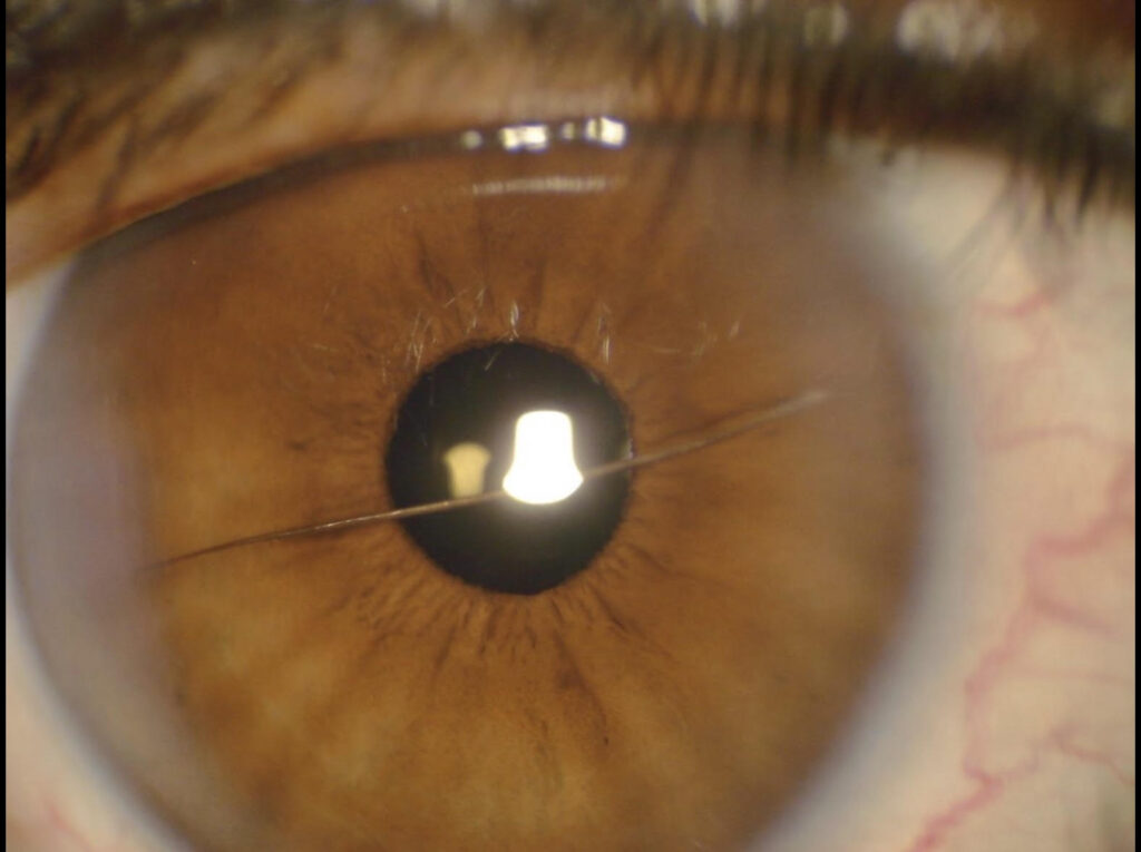
After confirming there is no evidence of penetration, topical anesthesia allows comfortable removal, typically performed at the slit lamp using a fine needle, spud, or foreign body loop, taking care to remove the fragment cleanly without unnecessarily enlarging the corneal defect.
A rust ring, if present, can often be removed at the same visit using a rotating burr, though some clinicians prefer to leave a central or deep rust ring to soften and partially resolve over a day or two before attempting complete removal, to minimize the risk of excessive tissue removal in a single pass.
This staged approach is particularly reasonable when the rust ring sits close to the visual axis, since a more conservative, patient approach there reduces the chance of leaving a visually significant residual scar from overly aggressive debridement.
After removal, treatment follows the same general principles as any traumatic corneal abrasion: topical antibiotic prophylaxis while the epithelial defect heals, cycloplegia for comfort if significant ciliary spasm or photophobia is present, and avoidance of a pressure patch in contaminated injuries (organic material, vegetation, contact lens–related injuries) given the increased infection risk in these specific scenarios.
Follow-Up and Complications
Most uncomplicated corneal foreign body injuries heal within a few days with no lasting visual consequence, though a follow-up exam is reasonable to confirm complete epithelial healing and to check for any residual rust staining or early signs of infection.
Risk of secondary microbial keratitis is elevated with any corneal epithelial defect, which is part of why routine antibiotic prophylaxis is standard during the healing period rather than reserved only for higher-risk injuries.
Organic or vegetative foreign bodies carry a specific additional risk of fungal infection that warrants closer follow-up than a typical inorganic foreign body from metal or glass, and patients with this exposure history should be specifically counseled to return promptly if symptoms worsen rather than improve as expected.


Document what you see
Two smartphone imaging tools built for everyday clinic use — one for the slit lamp, one for the fundus.
From Choroida — the team behind this siteReferences
- Bagheri N, Wajda B, eds. The Wills Eye Manual.
- Kuhn F, Morris R, Witherspoon CD, Mann L. Epidemiology of blinding trauma in the United States Eye Injury Registry. Ophthalmic Epidemiology.
- American Academy of Ophthalmology. Basic and Clinical Science Course, Section 8: External Disease and Cornea.
Test yourself
A few questions straight from this article.
-
Which injury mechanism carries a genuine risk that a fragment has penetrated the globe?
High-velocity mechanisms such as hammering, grinding and drilling can drive a fragment into the globe, whereas low-velocity mechanisms carry minimal penetration risk. -
Why can a penetrating high-velocity metallic injury be overlooked on examination?
A fragment may lodge intraocularly through an entry wound so small it is easily missed, which is why a metal-on-metal history should prompt a deliberate search for penetration. -
A brownish rust ring around a corneal foreign body indicates what?
A rust ring forms when an iron- or steel-containing fragment has been present long enough for oxidation to begin, appearing as brown discoloration around the entry site. -
Which finding indicates full-thickness penetration rather than a superficial corneal foreign body?
A shallow or irregular anterior chamber, a peaked or teardrop pupil, a positive Seidel test or visible intraocular contents turn a routine removal into a surgical emergency. -
Which imaging study is used when an intraocular metallic foreign body is suspected?
CT of the orbit is used to look for an intraocular or intraorbital foreign body once penetration is suspected, alongside urgent ophthalmology involvement. -
Why is MRI contraindicated when a metallic intraocular foreign body is suspected?
A metallic fragment can be moved magnetically during scanning, so CT rather than MRI is used when metal is suspected. -
Which symptom pattern after an eye injury raises suspicion of globe penetration?
Pain that settles and then worsens can occur when a fragment penetrates and settles intraocularly, and belongs on the list of features prompting CT imaging. -
How is an uncomplicated corneal foreign body removed once penetration has been excluded?
Topical anaesthesia allows comfortable slit-lamp removal, taking care to lift the fragment cleanly without enlarging the corneal defect unnecessarily. -
Why might a deep or central rust ring be left to soften for a day or two?
Near the visual axis a staged approach is reasonable, since aggressive debridement in a single pass risks leaving a visually significant residual scar. -
In which corneal foreign body injuries should a pressure patch be avoided?
These contaminated injuries carry an increased infection risk, so a pressure patch is avoided while the epithelial defect heals under antibiotic cover.
