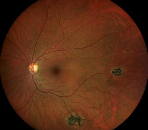
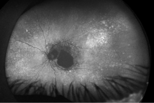
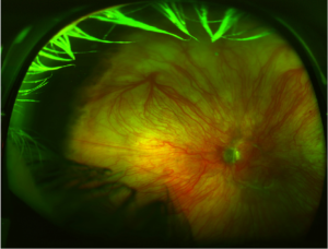
A single blow to the eye can tear the iris loose from its anchor point — and the visible defect is often the least important part of the injury.
Iridodialysis is traumatic separation of the iris root from the ciliary body, and it is a marker of significant blunt ocular trauma.
The disc-shaped gap it leaves at the iris periphery can look dramatic, distorting the pupil and letting light enter the eye through an extra opening.
What actually matters most, though, is what else the same blow did — hyphema, angle recession, lens subluxation, and retinal injury frequently travel together with iridodialysis.
Treating the visible iris defect while missing one of these associated injuries is a common and consequential mistake.
A systematic, complete examination after any blunt trauma severe enough to cause iridodialysis is what actually protects the eye.
What Is Iridodialysis?
Iridodialysis is a traumatic disinsertion of the iris root from its attachment at the ciliary body, following blunt ocular trauma.
Its defining features include:
- A crescentic or disc-shaped defect at the peripheral iris, often visible as a dark gap adjacent to the limbus
- Distortion of the pupil shape (corectopia) when the defect is large
- Frequent coexistence with other blunt trauma findings — hyphema, angle recession, and lens subluxation among them
- A range of severity from a small, asymptomatic defect to a large dialysis causing significant glare and monocular diplopia
Iridodialysis is best understood as one visible marker of a blunt trauma event, not an isolated injury — the mechanism that tore the iris root frequently damaged other structures too.
Epidemiology
Iridodialysis occurs in the setting of significant blunt ocular trauma across a range of settings.
- Common mechanisms include sports injuries (particularly from balls or similar projectiles), motor vehicle collisions, fistfights, and airbag deployment
- It is more common in young, active individuals, reflecting the typical demographic for blunt ocular trauma generally
- Frequently occurs alongside hyphema and angle recession, since all three result from the same mechanical force distributed through the anterior segment
Because the same blunt force mechanism produces a cluster of related injuries, finding an iridodialysis should prompt a deliberate search for its usual companions rather than being treated as an isolated finding.
All-fit Slit-Lamp Adapter
Record and share exactly what you see at the slit lamp. One adapter fits any slit lamp or surgical microscope — and any smartphone.
From Choroida — the team behind this sitePathophysiology
The injury results from sudden mechanical deformation of the globe at the moment of impact.
- Blunt force causes rapid anteroposterior compression and equatorial expansion of the globe
- This sudden deformation places shearing stress on the iris root, its thinnest and most mechanically vulnerable point of attachment
- Sufficient stress tears the iris root away from the ciliary body, producing the dialysis
- The same compressive-expansive force commonly damages the adjacent anterior chamber angle (angle recession) and can rupture iris or ciliary body vessels, producing hyphema
- More severe trauma can extend the same mechanical stress to zonular fibers, causing lens subluxation, or posteriorly to the retina and choroid
Because a single mechanical event drives all of these findings simultaneously, the extent of iridodialysis often serves as a rough index of how much force the eye actually absorbed.
Risk Factors
Mechanism-Related Risk Factors
- Sports involving a projectile capable of striking the orbit (racquet sports, baseball, and similar)
- Motor vehicle collisions, including airbag-related trauma
- Interpersonal violence and assault
- Occupational blunt trauma exposure
Patient-Related Risk Factors
- Young, active age groups, given typical exposure to high-risk activities
- Lack of protective eyewear during high-risk sports
Protective eyewear during high-risk sports remains one of the few genuinely effective preventive measures against this entire cluster of blunt trauma injuries.
Clinical Presentation
Symptoms
- A history of recent blunt trauma to the eye or orbit
- Blurred vision, which may reflect the dialysis itself, an associated hyphema, or other concurrent injury
- Glare or monocular diplopia when a large dialysis creates a second functional opening for light to enter the eye
- Eye pain, tearing, and photophobia in the acute setting
Examination Findings
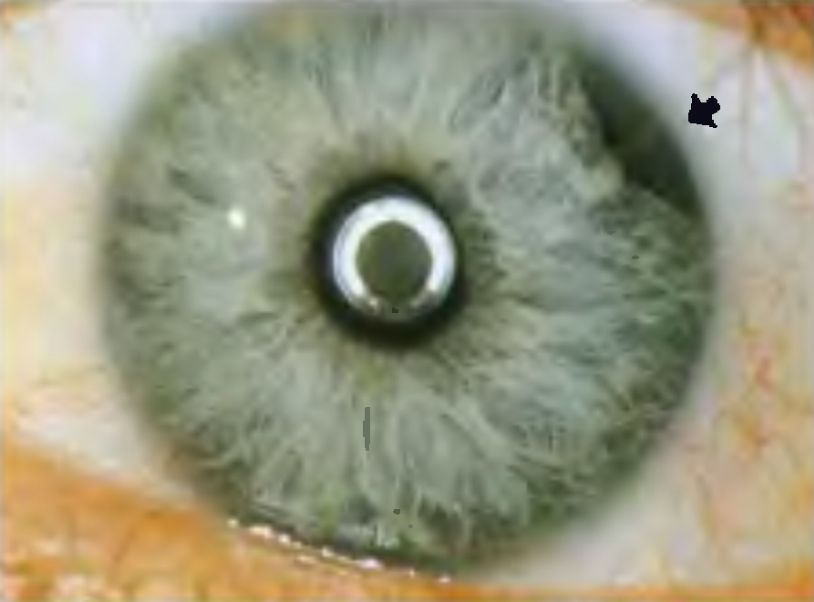
- A crescentic or disc-shaped defect at the iris root, visible as a dark gap between the iris periphery and the limbus
- Distortion of pupil shape when the defect is significant
- Associated findings on careful exam — hyphema, angle recession on gonioscopy, lens subluxation, or vitreous hemorrhage
- A dilated fundus exam is essential to look for commotio retinae, retinal breaks, or choroidal rupture from the same trauma
Every patient with iridodialysis needs a complete anterior and posterior segment exam, including gonioscopy once any hyphema allows it — the iris defect itself is rarely the only injury present.
Diagnostic Evaluation
Slit-Lamp Examination
- Detailed assessment of the iris defect size, location, and any associated anterior chamber findings
- Intraocular pressure measurement, since both acute hyphema and, later, angle recession can affect IOP
Gonioscopy
- Performed once any hyphema has settled sufficiently, to assess for angle recession, a common companion injury with long-term glaucoma risk
Dilated Fundus Examination and Imaging
- Careful examination for commotio retinae, retinal breaks, choroidal rupture, or vitreous hemorrhage
- B-scan ultrasonography when the view is obscured by hyphema or vitreous hemorrhage
- Ultrasound biomicroscopy can further characterize the dialysis and any associated ciliary body or zonular injury when needed
Angle recession found on gonioscopy carries a lifelong glaucoma risk, which is why every iridodialysis patient needs long-term IOP monitoring, not just acute-phase care.
Differential Diagnosis
The traumatic history usually makes the diagnosis clear, but consider distinguishing from:
- Coloboma — a congenital iris defect, typically inferior and present since birth, without a trauma history
- Surgical iridectomy — a clean, typically peripheral defect with a surgical history rather than trauma
- Iris tumor with associated pupil distortion — a mass lesion rather than a true structural defect at the root
A clear history of blunt trauma, combined with the classic peripheral location at the iris root, distinguishes iridodialysis from these mimics without much difficulty.
Management
Observation
- Appropriate for small, asymptomatic dialyses without significant visual symptoms
- Concurrent management of any associated hyphema, elevated IOP, or other trauma findings takes priority in the acute phase
Surgical Repair
- Indicated for large dialyses causing significant glare, monocular diplopia, or cosmetic concern
- Surgical iridodialysis repair (iridopexy) reattaches the iris root, typically using sutures passed through the peripheral iris and secured externally or internally
- Combined management is often needed when lens subluxation or other concurrent injury requires its own surgical correction
Long-Term Monitoring
- Ongoing IOP surveillance given the elevated lifetime glaucoma risk associated with angle recession, which can manifest years after the initial injury
- Regular fundus examination given the increased risk of retinal complications after significant blunt trauma
Surgical repair addresses the visible defect, but the long-term risk that actually matters most — angle recession glaucoma — requires monitoring that continues long after the eye looks fully healed.
Prognosis
Visual prognosis depends far more on associated injuries than on the iridodialysis itself.
- An isolated, small iridodialysis without other significant trauma findings carries an excellent visual prognosis
- Concurrent hyphema, lens subluxation, or posterior segment injury can significantly worsen overall visual outcome
- Angle recession, when present, carries a lifelong risk of secondary glaucoma that can develop even decades after the original injury
Long-term follow-up after iridodialysis is not optional — the glaucoma risk from an associated angle recession can silently develop long after the patient has forgotten about the original injury.


Document what you see
Two smartphone imaging tools built for everyday clinic use — one for the slit lamp, one for the fundus.
From Choroida — the team behind this siteReferences
- Kaufman JH, Tolpin DW. Glaucoma after traumatic angle recession: a ten-year prospective study. American Journal of Ophthalmology. 1974.
- Ashaye AO. Traumatic iridodialysis at the University College Hospital, Ibadan. West African Journal of Medicine. 2000.
- Iridodialysis. StatPearls, NCBI Bookshelf.
- Ocular Blunt Trauma. EyeWiki, American Academy of Ophthalmology.
- Sihota R, Sood NN, Agarwal HC. Traumatic glaucoma: a prospective study. Acta Ophthalmologica Scandinavica. 1995.
Test yourself
A few questions straight from this article.
-
Iridodialysis is defined as traumatic separation of the iris root from which structure?
Iridodialysis is a traumatic disinsertion of the iris root from its attachment at the ciliary body after blunt ocular trauma. -
Which mechanical event at impact tears the iris root in iridodialysis?
Blunt force compresses the globe anteroposteriorly while expanding it at the equator, and that sudden deformation shears the iris away from the ciliary body. -
Why does the iris root give way first in blunt ocular trauma causing iridodialysis?
Shearing stress concentrates at the iris root because it is the thinnest and most mechanically vulnerable point of iris attachment. -
A large iridodialysis characteristically produces which pair of visual symptoms?
A large dialysis creates a second functional opening for light to enter the eye, which the patient experiences as glare and monocular double vision. -
What is the classic slit-lamp appearance of iridodialysis?
The defect appears as a crescentic or disc-shaped dark gap between the iris periphery and the limbus, often with pupil distortion when large. -
In iridodialysis, when is gonioscopy performed and what is it looking for?
Gonioscopy is deferred until any hyphema has settled sufficiently, then used to look for angle recession, the companion injury that carries long-term glaucoma risk. -
Which test is used in iridodialysis when hyphema or vitreous hemorrhage obscures the fundus view?
B-scan ultrasonography evaluates the posterior segment when the media are opaque; ultrasound biomicroscopy can additionally characterise the dialysis and any ciliary body or zonular injury. -
Which feature best distinguishes iridodialysis from a congenital iris coloboma?
Coloboma is congenital and typically inferior with no trauma history, whereas iridodialysis follows blunt trauma and sits classically at the iris root. -
Which iridodialysis warrants surgical repair (iridopexy) rather than simple observation?
Small asymptomatic defects are observed; repair with sutures reattaching the iris root is reserved for large dialyses causing glare, monocular diplopia or cosmetic concern. -
Which long-term complication makes lifelong follow-up mandatory after iridodialysis?
Angle recession carries a lifelong glaucoma risk that can surface decades after the injury, so intraocular pressure surveillance must continue long after the eye looks healed.

Writes and reviews the clinical articles on Educate. Every post is checked against peer-reviewed sources before it goes live.