
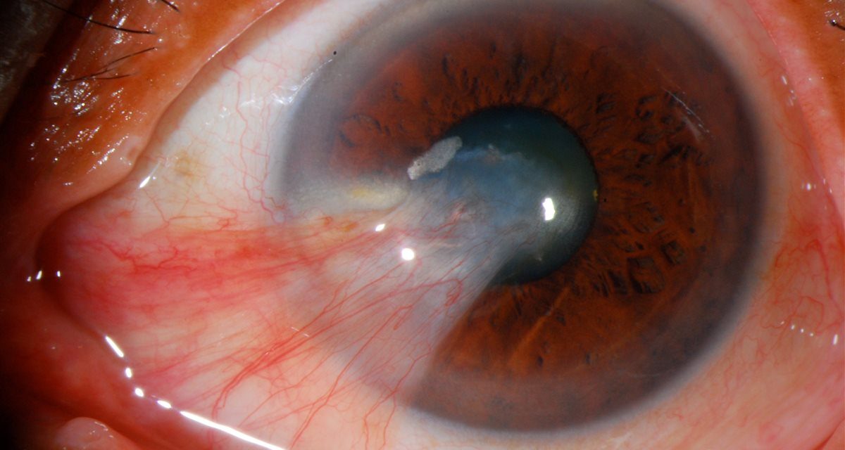
Most eyes with a pterygium have lived in bright sun.
The lesion looks simple: a wedge of pink tissue creeping in from the corner of the eye. Yet it is often dismissed as cosmetic until it starts to blur vision, pull on the cornea, or come back after surgery.
A pterygium is one of the most frequent reasons a patient with a “red, growing spot” walks into clinic. Reading it correctly — and separating it from the lesions it mimics — is a core anterior-segment skill.
What Is a Pterygium?
A pterygium is a wing-shaped fibrovascular growth of the bulbar conjunctiva that extends across the limbus onto the cornea. The name comes from the Greek pterygion, meaning “little wing.”
It has three recognizable parts: a head (the leading apex on the cornea), a neck (at the limbus), and a body (the conjunctival portion). A faint iron line — the Stocker line — may sit in the corneal epithelium just ahead of the advancing head.
The overwhelming majority are nasal, though temporal and double (nasal plus temporal) pterygia occur. Understanding that a pterygium is true fibrovascular tissue — not simply a deposit — is what explains its tendency to distort the cornea and recur.
Epidemiology and Risk Factors
Pterygium is strongly a disease of environment. Prevalence rises sharply near the equator and in populations with heavy outdoor exposure, giving rise to the description of a “pterygium belt” in latitudes closer to the tropics.
- Ultraviolet light is the dominant risk factor — the reason outdoor workers, fishermen, and farmers are over-represented.
- Chronic surface irritation from wind, dust, and dryness contributes.
- Age and male sex are associated, largely through cumulative sun exposure.
- Nasal predominance is thought to reflect light focused onto the nasal limbus after entering from the temporal side.
The practical message is that a pterygium is, in part, a marker of a lifetime of unprotected sun — which is why prevention advice matters as much as the lesion itself.
All-fit Slit-Lamp Adapter
Record and share exactly what you see at the slit lamp. One adapter fits any slit lamp or surgical microscope — and any smartphone.
From Choroida — the team behind this sitePathogenesis
Chronic ultraviolet exposure damages the limbal stem-cell barrier and drives a cascade of changes in the conjunctival tissue: activation of altered fibroblasts, new vessel formation, connective-tissue breakdown (elastotic degeneration), and inflammation.
The result is an actively growing, invasive-looking tissue that advances onto the cornea by dissolving and replacing the normal architecture at the leading edge. This is why a pterygium behaves less like a passive plaque and more like a slowly proliferating growth — and why the same biology fuels recurrence after removal.
Clinical Presentation
Symptoms range from none to significant, and often do not match the lesion’s size.
- Irritation, redness, and a foreign-body sensation, worse in wind and dry conditions.
- Cosmetic concern — frequently the main complaint.
- Blurred vision from induced astigmatism as the lesion flattens the cornea, or from the head encroaching on the visual axis.
- Intermittent inflammation, with episodes of a redder, more elevated, and more vascular lesion.
- Restricted motility or diplopia in advanced or recurrent cases with fibrosis.
On slit-lamp examination the pterygium is a triangular, vascularized fold with its apex on the cornea; documenting how far the head has crossed the limbus toward the pupil is the single most useful measurement to record over time.
Why It Is So Often Underestimated
Two errors are common. The first is treating every pterygium as purely cosmetic; a lesion approaching the visual axis or inducing progressive astigmatism is a functional problem, not a vanity one.
The second is confusing it with lesions that share the same corner of the eye. A pinguécula looks similar but stops at the limbus and never crosses onto the cornea. More importantly, an atypical, thickened, or leukoplakic “pterygium” — especially one that is unusually elevated, gelatinous, or temporal — can be ocular surface squamous neoplasia (OSSN) in disguise.
The safe habit is to treat any pterygium that looks atypical as a lesion to be examined carefully, and biopsied when excised, rather than assumed benign.
Diagnostic Evaluation
The diagnosis is clinical, but a few steps sharpen management.
- Slit-lamp assessment of the head, extent onto the cornea, vascularity, and any involvement of the visual axis.
- Refraction and keratometry / corneal topography to quantify induced astigmatism — often the objective reason to operate.
- Serial photography and measurement to document progression.
- Histopathology of any excised or atypical lesion to exclude OSSN.
Topography, in particular, frequently reveals more visual impact than the naked-eye appearance suggests.
Differential Diagnosis
- Pinguecula — similar tissue that does not cross the limbus.
- Pseudopterygium — a fold of conjunctiva adherent to the cornea after prior injury, chemical burn, or inflammation; unlike a true pterygium, a probe can usually be passed beneath its neck.
- Ocular surface squamous neoplasia — the diagnosis not to miss.
- Limbal dermoid and other congenital lesions.
- Nodular episcleritis or a conjunctival granuloma in inflamed cases.
Distinguishing a true pterygium from a pseudopterygium at the slit lamp changes both the workup and the surgical approach.
Management
Most quiet pterygia need only reassurance and surface care.
- Conservative measures: lubricating drops for irritation, short courses of a mild topical anti-inflammatory during flares, and — crucially — ultraviolet protection with wraparound sunglasses and a hat to slow progression.
- Surgical excision is indicated for a lesion encroaching on the visual axis, causing significant astigmatism, limiting motility, recurrently inflamed, or cosmetically unacceptable to the patient.
- Technique matters more than the decision to operate. Bare-sclera excision alone carries a high recurrence rate, so the standard of care pairs excision with conjunctival or limbal-conjunctival autograft (often secured with tissue adhesive rather than sutures). Amniotic membrane and adjuncts such as mitomycin C are used in selected or recurrent cases.
The goal of surgery is not just removal but a low-recurrence closure, which is why the reconstruction — not the excision — defines a good result.
Prognosis and Recurrence
The visual and cosmetic outcome after well-performed autograft surgery is generally excellent. The main challenge is recurrence, which tends to appear within the first months to a year and is more likely in younger patients, fleshy and vascular lesions, and after bare-sclera technique.
A recurrent pterygium is typically more aggressive and more fibrotic than the original, so choosing a low-recurrence technique and reinforcing sun protection at the first operation is the best long-term strategy.
Would you like to document anterior-segment lesions like pterygium with your smartphone?


Document what you see
Two smartphone imaging tools built for everyday clinic use — one for the slit lamp, one for the fundus.
From Choroida — the team behind this siteReferences
- Chui J, Coroneo MT, Tat LT, et al. Ophthalmic pterygium: a stem cell disorder with premalignant features. American Journal of Pathology. 2011;178(2):817-827.
- Liu L, Wu J, Geng J, et al. Geographical prevalence and risk factors for pterygium: a systematic review and meta-analysis. BMJ Open. 2013;3(11):e003787.
- Clearfield E, Muthappan V, Wang X, Kuo IC. Conjunctival autograft for pterygium. Cochrane Database of Systematic Reviews. 2016;(2):CD011349.
- Kaufman SC, Jacobs DS, Lee WB, et al. Options and adjuvants in surgery for pterygium: a report by the American Academy of Ophthalmology. Ophthalmology. 2013;120(1):201-208.
Test yourself
A few questions straight from this article.
-
Which description best defines a pterygium?
A pterygium is a wing-shaped fibrovascular growth of bulbar conjunctiva that extends across the limbus onto the cornea, with a head on the cornea, a neck at the limbus and a conjunctival body. -
The Stocker line associated with a pterygium is which finding?
A faint iron line, the Stocker line, may sit in the corneal epithelium just ahead of the advancing head of the pterygium. -
Which factor is the dominant risk factor for developing a pterygium?
Ultraviolet light is the dominant risk factor, which is why outdoor workers, fishermen and farmers are over-represented and why prevalence rises near the equator. -
In the pathogenesis of pterygium, chronic ultraviolet exposure damages which structure?
Ultraviolet damage to the limbal stem-cell barrier triggers altered fibroblasts, new vessel formation, elastotic degeneration and inflammation, producing a tissue that invades the cornea and tends to recur. -
What distinguishes a pinguecula from a pterygium at the slit lamp?
A pinguecula looks like similar tissue but stops at the limbus, whereas a true pterygium extends across the limbus onto the cornea. -
Which slit-lamp finding suggests a pseudopterygium rather than a true pterygium?
A pseudopterygium is a conjunctival fold stuck to the cornea after injury, chemical burn or inflammation, and unlike a true pterygium a probe can usually be passed under its neck. -
An unusually elevated, gelatinous or leukoplakic pterygium-like lesion should raise suspicion for which diagnosis?
An atypical, thickened, leukoplakic or temporal lesion can be ocular surface squamous neoplasia in disguise, so any atypical pterygium should be sent for histopathology when excised. -
Which investigation best quantifies the visual impact of a pterygium?
Refraction with keratometry or topography quantifies the induced astigmatism, often revealing more visual impact than the naked-eye appearance suggests, and is frequently the objective reason to operate. -
Which surgical approach is the standard of care for pterygium because of recurrence risk?
Bare-sclera excision alone carries a high recurrence rate, so excision is paired with a conjunctival or limbal-conjunctival autograft, often secured with tissue adhesive; amniotic membrane and mitomycin C are reserved for selected or recurrent cases. -
When does recurrence after pterygium surgery typically appear?
Recurrence tends to appear within the first months to a year and is more likely in younger patients, fleshy vascular lesions and after bare-sclera technique; a recurrent pterygium is usually more aggressive and fibrotic than the original.
