
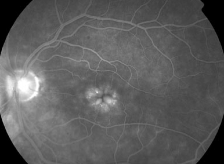
Fluorescein angiography (FA) remains one of the most valuable imaging modalities in retinal disease. One of the most common findings encountered during angiographic evaluation is vascular leakage, which often manifests as progressive hyperfluorescence in the late phases of the study.


For many ophthalmologists, especially when evaluating patients with retinal edema or microvascular abnormalities, vascular leakage immediately raises suspicion for diabetic retinopathy.
While diabetic retinal disease is undoubtedly a major cause of angiographic leakage, it is far from the only one.
Numerous retinal, inflammatory, vascular, infectious, and neoplastic disorders can produce leaking vessels on angiography.
Misinterpreting every case of vascular leakage as diabetic retinopathy can delay diagnosis, lead to inappropriate treatment, and potentially overlook sight-threatening systemic diseases.
For retina specialists, angiographic leakage should be viewed as a finding—not a diagnosis.
Understanding Vascular Leakage on Fluorescein Angiography
Leakage occurs when the blood-retinal barrier becomes disrupted, allowing fluorescein dye to escape from retinal or choroidal vessels into surrounding tissues.
This process appears as:
- Progressive hyperfluorescence
- Increasing lesion size over time
- Blurring of vascular margins
- Diffuse retinal staining in late frames
Leakage indicates vascular dysfunction but does not identify the underlying cause.
👉 The critical question is not whether leakage exists, but why it exists.
Why Leakage Is Often Mistaken for Diabetic Retinopathy
Diabetic retinopathy is among the most common causes of retinal vascular leakage.
Typical angiographic findings include:
- Microaneurysm leakage
- Diffuse capillary leakage
- Macular edema
- Areas of capillary non-perfusion
- Neovascular leakage
Because diabetic retinopathy is so prevalent, clinicians may sometimes develop a diagnostic bias when interpreting angiograms.
However, several other diseases can produce remarkably similar findings.
1. Retinal Vein Occlusion
Both branch retinal vein occlusion (BRVO) and central retinal vein occlusion (CRVO) frequently cause significant leakage.
Angiographic Features
- Dilated tortuous vessels
- Diffuse capillary leakage
- Macular edema
- Areas of ischemia
Unlike diabetic retinopathy, the leakage often follows a specific vascular distribution corresponding to the affected vein.
Clinical Clues
- Sudden visual loss
- Sectoral retinal hemorrhages (BRVO)
- “Blood-and-thunder” appearance (CRVO)
👉 Leakage confined to a venous territory should raise suspicion for retinal vein occlusion.
2. Retinal Vasculitis
Inflammatory vascular disease is an important non-diabetic cause of leakage.
Angiographic Findings
- Vessel wall staining
- Perivascular leakage
- Diffuse capillary leakage
- Optic disc leakage
Common Causes
- Sarcoidosis
- Behçet disease
- Tuberculosis
- Systemic lupus erythematosus
- Idiopathic retinal vasculitis
Unlike diabetic retinopathy, leakage often involves vessel walls themselves rather than isolated microaneurysms.
👉 Extensive vascular leakage in a young patient should always prompt consideration of vasculitis.
3. Uveitic Macular Edema
Inflammatory diseases frequently produce angiographic leakage.
Typical Findings
- Diffuse retinal leakage
- Cystoid macular edema
- Disc hyperfluorescence
- Peripheral vascular leakage
Common associated conditions include:
- Intermediate uveitis
- Birdshot chorioretinopathy
- Behçet disease
- Sarcoidosis
In some cases, leakage may be far more extensive than the clinical examination suggests.
4. Radiation Retinopathy
Radiation-induced microvascular damage can closely mimic diabetic retinopathy.
Angiographic Features
- Capillary leakage
- Microaneurysms
- Macular edema
- Capillary dropout
Important Historical Clue
A prior history of:
- Plaque brachytherapy
- External beam radiation
- Orbital radiation treatment
may be the key to diagnosis.
👉 Radiation retinopathy is often called “diabetic retinopathy without diabetes.”
5. Ocular Ischemic Syndrome
Chronic ocular hypoperfusion may produce significant vascular leakage.
Fluorescein Angiography Findings
- Delayed choroidal filling
- Delayed arterial filling
- Mid-peripheral leakage
- Prolonged arteriovenous transit time
Clinical Clues
- Carotid artery disease
- Ocular pain
- Neovascularization
- Asymmetric retinal findings
The delayed filling pattern often distinguishes it from diabetic disease.
6. Retinal Telangiectatic Disorders
Several vascular disorders produce leakage through abnormal retinal capillaries.
Examples
- Macular telangiectasia type 1
- Macular telangiectasia type 2
- Coats disease
- Leber miliary aneurysms
Angiographic Features
- Telangiectatic vessels
- Focal leakage
- Lipid exudation
The distribution and morphology of abnormal vessels often provide important diagnostic clues.
7. Neovascular Age-Related Macular Degeneration
Leakage originating from choroidal neovascularization can sometimes be confused with retinal vascular leakage.
FA Findings
- Classic leakage patterns
- Occult leakage
- Late staining
OCT Correlation
Often reveals:
- Subretinal fluid
- Pigment epithelial detachments
- Intraretinal cysts
👉 Not all macular leakage originates from retinal vessels.
8. Central Serous Chorioretinopathy (CSC)
CSC represents another important mimic.
Characteristic Findings
- Ink-blot leakage
- Smoke-stack leakage
- Focal retinal pigment epithelial defects
Unlike diabetic retinopathy, leakage originates from the retinal pigment epithelium rather than retinal capillaries.
9. Retinal Capillary Hemangioblastoma
Vascular tumors may demonstrate dramatic leakage on angiography.
Features Include
- Early hyperfluorescence
- Progressive leakage
- Dilated feeder vessels
These lesions may occur sporadically or as part of Von Hippel-Lindau Disease.
10. Infectious Retinitis
Several infectious diseases can cause vascular leakage.
Examples
- Ocular toxoplasmosis
- Syphilitic retinitis
- Tuberculous retinal vasculitis
- Viral retinitis
Angiographic Findings
- Perivascular leakage
- Disc leakage
- Areas of active inflammation
Infectious etiologies should always be considered when leakage is accompanied by inflammatory signs.
The Importance of Wide-Field Angiography
Ultra-widefield angiography has expanded our understanding of peripheral retinal disease.
It may reveal:
- Peripheral vasculitis
- Peripheral ischemia
- Neovascularization
- Capillary non-perfusion
Many conditions initially thought to be isolated macular disease are now recognized as diffuse retinal disorders.
👉 Significant peripheral leakage may be present despite minimal posterior pole findings.
OCT and OCT Angiography: Essential Partners
Fluorescein angiography should rarely be interpreted in isolation.
OCT Helps Evaluate
- Macular edema
- Retinal architecture
- Intraretinal fluid
- Subretinal fluid
OCT Angiography Helps Identify
- Non-perfusion
- Neovascular complexes
- Capillary abnormalities
Combining modalities often improves diagnostic accuracy substantially.
Red Flags That Suggest “Not Diabetic Retinopathy”
Ophthalmologists should broaden their differential diagnosis when leakage is accompanied by:
- No history of diabetes
- Young patient age
- Significant vitreous inflammation
- Extensive vascular sheathing
- Delayed vascular filling
- Focal telangiectatic lesions
- Choroidal abnormalities
- Asymmetric involvement
👉 When the clinical picture does not fit diabetic retinopathy, the angiogram deserves a second look.
A Practical Diagnostic Approach
When encountering leaking vessels on angiography:
Step 1
Assess the pattern of leakage.
Is it:
- Focal?
- Diffuse?
- Perivascular?
- Neovascular?
Step 2
Evaluate associated findings.
Look for:
- Ischemia
- Inflammation
- Disc leakage
- Choroidal abnormalities
Step 3
Correlate with clinical examination.
Step 4
Use OCT and OCT angiography.
Step 5
Consider systemic associations.
Many causes of leakage are linked to systemic inflammatory, vascular, or infectious diseases.
Would you have interest in taking retinal images with your smartphone?
Fundus photography is superior to fundus analysis as it enables intraocular pathologies to be photo-captured and encrypted information to be shared with colleagues and patients.
Recent technologies allow smartphone-based attachments and integrated lens adaptors to transform the smartphone into a portable fundus camera and Retinal imaging by smartphone.
RETINAL IMAGING BY YOUR SMARTPHONE
References
- Yannuzzi LA, et al. Retina Atlas. Elsevier.
- Spaide RF, Curcio CA. “Anatomical correlates to angiographic patterns in retinal disease.” Retina. 2010.
- Campochiaro PA. “Retinal vascular diseases and fluorescein angiography.” Ophthalmology. 2015.
- Nicholson BP, Schachat AP. “A review of clinical trials of anti-VEGF agents for retinal vascular diseases.” Retina. 2010.
- Pichi F, et al. “Wide-field imaging in retinal vasculitis.” Prog Retin Eye Res. 2020.
RETINAL IMAGING BY YOUR SMARTPHONE

RETINAL IMAGING BY YOUR SMARTPHONE