
Demyelinating optic neuropathy is one of the most important causes of acute optic nerve dysfunction in young and middle-aged adults.

For decades, optic neuritis has been closely associated with multiple sclerosis (MS), and many ophthalmologists instinctively consider MS when evaluating a patient with painful visual loss and optic nerve inflammation.
However, modern neuro-ophthalmology has revealed that demyelinating optic neuropathy extends far beyond multiple sclerosis.
A growing number of inflammatory and autoimmune disorders can affect the optic nerve through demyelinating mechanisms, often presenting with distinct clinical features, prognoses, and treatment strategies.
Recognizing these alternative causes is critical because some conditions carry a higher risk of severe visual loss, frequent relapses, and systemic neurological complications if left untreated.
For ophthalmologists, understanding the broader spectrum of demyelinating optic neuropathies has become increasingly important in the era of targeted immunotherapy.
What Is Demyelinating Optic Neuropathy?
Demyelinating optic neuropathy refers to optic nerve dysfunction resulting from inflammatory damage to the myelin sheath surrounding optic nerve axons.
This process disrupts normal neural conduction and may lead to:
- Acute visual loss
- Color vision impairment
- Visual field defects
- Relative afferent pupillary defect (RAPD)
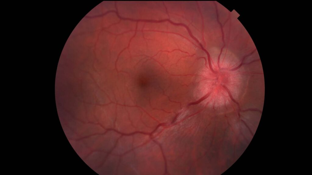
- Optic disc edema or retrobulbar optic neuritis
Although multiple sclerosis remains a major cause, it is no longer considered the only demyelinating disease affecting the optic nerve.
Why Looking Beyond Multiple Sclerosis Matters
Historically, many cases of optic neuritis were classified simply as “MS-related.”
Today, several distinct entities have been identified, including:
- Multiple sclerosis-associated optic neuritis
- Neuromyelitis optica spectrum disorder (NMOSD)
- Myelin oligodendrocyte glycoprotein antibody-associated disease (MOGAD)
- Acute disseminated encephalomyelitis (ADEM)
- Other autoimmune and inflammatory demyelinating disorders
👉 These conditions may appear similar initially but differ dramatically in prognosis and management.
Classic Multiple Sclerosis-Associated Optic Neuritis
MS-related optic neuritis remains the most familiar form.
Typical Features
- Young adults (often 20–45 years)
- Unilateral involvement
- Pain with eye movement
- Moderate visual loss
- Good spontaneous recovery
- Retrobulbar involvement common
Patients frequently report:
- Decreased color perception
- Central scotoma
- Reduced contrast sensitivity
MRI often demonstrates demyelinating brain lesions suggestive of multiple sclerosis.
👉 The classic presentation of painful unilateral visual loss remains highly suggestive of MS-associated optic neuritis.
Neuromyelitis Optica Spectrum Disorder (NMOSD)
NMOSD is an autoimmune astrocytopathy associated with antibodies against aquaporin-4 (AQP4).
Unlike MS, NMOSD often produces more severe optic nerve injury.
Clinical Clues
- Severe visual loss
- Bilateral involvement
- Poor spontaneous recovery
- Frequent relapses
- Extensive optic nerve lesions on MRI
Many patients present with vision reduced to counting fingers, hand motion, or worse.
MRI Findings
Often reveal:
- Long-segment optic nerve involvement
- Chiasmal extension
- Extensive inflammatory lesions
👉 Severe optic neuritis with poor recovery should immediately raise suspicion for NMOSD.
Myelin Oligodendrocyte Glycoprotein Antibody Disease (MOGAD)
MOG antibody-associated disease has emerged as a distinct demyelinating disorder over the past decade.
Its optic neuritis often differs from both MS and NMOSD.
Characteristic Features
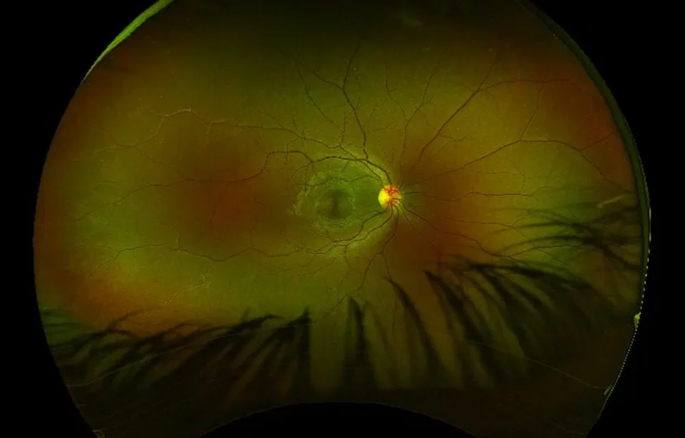
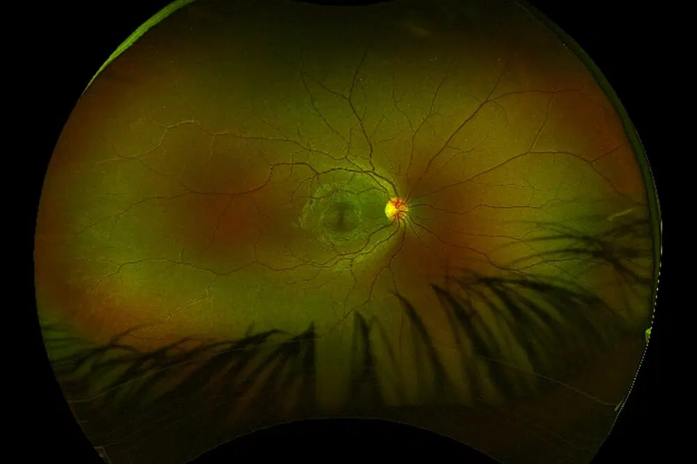
- Marked optic disc edema
- Severe acute visual loss
- Bilateral involvement more common
- Excellent recovery after treatment
- Frequent recurrence
Many patients show dramatic disc swelling that appears disproportionate to visual dysfunction.
MRI Characteristics
Often demonstrate:
- Anterior optic nerve involvement
- Perineural enhancement
- Long-segment inflammation
👉 Prominent optic disc edema is one of the most useful clues suggesting MOG-associated optic neuritis.
Acute Disseminated Encephalomyelitis (ADEM)
ADEM is an inflammatory demyelinating disorder that typically occurs after infections or vaccinations, particularly in children.
Clinical Features
- Encephalopathy
- Multifocal neurological deficits
- Bilateral optic neuritis
- Brain MRI abnormalities
Optic nerve involvement may occur simultaneously with widespread central nervous system inflammation.
Clinical Features That Suggest an Atypical Demyelinating Optic Neuropathy
Not all optic neuritis behaves like classic MS-related disease.
Certain findings should prompt further investigation.
Red Flags
- Bilateral visual loss
- Severe visual impairment
- Recurrent optic neuritis
- Poor visual recovery
- Extensive optic disc swelling
- Chiasmal involvement
- Severe retinal nerve fiber layer loss
👉 The more atypical the presentation, the broader the differential diagnosis should become.
The Role of MRI
MRI remains one of the most important diagnostic tools.
Brain MRI
Helps identify:
- Demyelinating plaques
- White matter lesions
- Alternative diagnoses
Orbital MRI
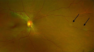
Can reveal:
- Optic nerve enhancement
- Lesion length
- Chiasmal involvement
- Perineural inflammation
Patterns of involvement often help distinguish MS, NMOSD, and MOGAD.
The Importance of Antibody Testing
The discovery of specific antibodies has transformed diagnosis.
Aquaporin-4 Antibody
Associated with:
- NMOSD
- Higher relapse risk
- Worse visual prognosis
MOG Antibody
Associated with:
- MOGAD
- Better visual recovery
- Frequent relapses
👉 Modern antibody testing has fundamentally changed the classification of optic neuritis.
Optical Coherence Tomography (OCT)
OCT provides valuable structural information following optic neuritis.
Findings May Include
- Retinal nerve fiber layer thinning
- Ganglion cell complex loss
- Optic nerve atrophy
Different diseases may produce varying degrees of retinal damage.
NMOSD often results in more profound structural loss than MS-associated optic neuritis.
Visual Field Patterns
Visual field defects vary considerably.
Common abnormalities include:
- Central scotomas
- Cecocentral defects
- Diffuse depression
- Altitudinal defects
- Arcuate defects
Although no pattern is completely disease-specific, severe diffuse defects are often seen in NMOSD.
Treatment Considerations
Acute Management
High-dose intravenous corticosteroids remain the standard first-line therapy.
Typical regimen:
- Intravenous methylprednisolone for several days
- Followed by oral taper in selected cases
Plasma Exchange (PLEX)
Particularly valuable in:
- NMOSD
- Steroid-refractory optic neuritis
- Severe visual loss
Early initiation may significantly improve outcomes.
Long-Term Management
Disease-specific therapy is essential.
Multiple Sclerosis
Treatment may involve:
- Disease-modifying therapies
- Neurological monitoring
- Relapse prevention
NMOSD
Requires aggressive immunosuppression because relapses often cause cumulative disability.
MOGAD
Management strategies continue to evolve but frequently involve immunotherapy for recurrent disease.
👉 Correct diagnosis directly influences long-term treatment decisions.
Prognosis
Visual outcomes vary considerably.
Best Prognosis
Often seen in:
- Typical MS-related optic neuritis
- Many MOG-associated cases
Worse Prognosis
Commonly associated with:
- NMOSD
- Recurrent attacks
- Delayed treatment
Repeated episodes can result in irreversible optic nerve atrophy and permanent visual impairment.
Future Perspectives
Advances in neuroimmunology continue to reshape the understanding of demyelinating optic neuropathies.
Emerging developments include:
- Novel biomarker discovery
- Targeted biological therapies
- Advanced MRI techniques
- Artificial intelligence-assisted imaging analysis
These innovations may improve diagnostic accuracy and enable earlier personalized treatment.
Would you have interest in taking retinal images with your smartphone?
Fundus photography is superior to fundus analysis as it enables intraocular pathologies to be photo-captured and encrypted information to be shared with colleagues and patients.
Recent technologies allow smartphone-based attachments and integrated lens adaptors to transform the smartphone into a portable fundus camera and Retinal imaging by smartphone.
RETINAL IMAGING BY YOUR SMARTPHONE
References
- Optic Neuritis Study Group. “Multiple sclerosis risk after optic neuritis: final optic neuritis treatment trial follow-up.” Arch Neurol. 2008.
- Beck RW, et al. “A randomized controlled trial of corticosteroids in the treatment of acute optic neuritis.” N Engl J Med. 1992.
- Wingerchuk DM, et al. “International consensus diagnostic criteria for neuromyelitis optica spectrum disorders.” Neurology. 2015.
- Jarius S, Wildemann B. “The history of neuromyelitis optica.” J Neuroinflammation. 2013.
- Jurynczyk M, et al. “Clinical presentation and prognosis in MOG-antibody disease.” Brain. 2017.
- Chen JJ, Flanagan EP. “Optic neuritis and autoimmune optic neuropathies.” Continuum (Minneap Minn). 2019.
- Petzold A, et al. “Diagnosis and classification of optic neuritis.” Lancet Neurology. 2022.
RETINAL IMAGING BY YOUR SMARTPHONE

RETINAL IMAGING BY YOUR SMARTPHONE