
Case Presentation
A 72-year-old female presented with an acute onset of severe right eye pain, redness, and profound vision loss over the past 48 hours.

Her medical history included untreated cataracts for over a decade and no prior ocular surgeries or systemic comorbidities.
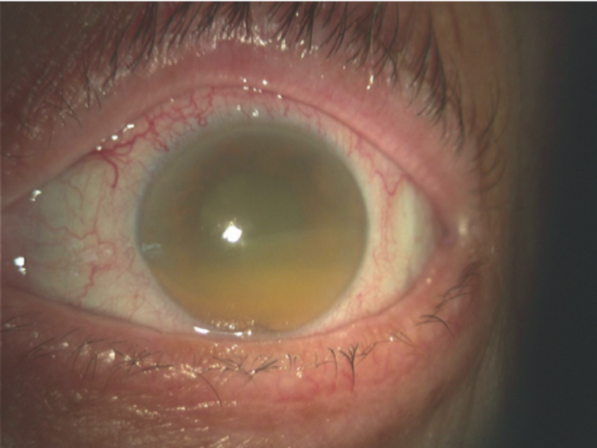
On examination, visual acuity was hand motion in the right eye and 20/30 in the left. Slit-lamp evaluation revealed a deep anterior chamber with 2+ flare, 3+ large translucent cells, and white flocculent material.
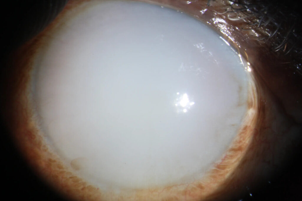
The right lens showed a hypermature cataract with a sunken sclerotic nucleus and phacodonesis.
Intraocular pressure measured 52 mmHg in the right eye via tonopen, with open angles on gonioscopy and mild corneal edema. A diagnosis of Phacolytic Glaucoma was made.
Disease Entity
Phacolytic glaucoma represents a secondary open-angle glaucoma arising from a hypermature cataract.
Ophthalmologists encounter this condition in patients with neglected cataracts, where lens protein leakage triggers acute intraocular pressure elevation.
Clinicians must recognize it promptly, as it threatens optic nerve integrity and corneal endothelium. Delayed intervention risks permanent vision loss.
This entity underscores the urgency of cataract surgery in advanced cases, distinguishing it from primary glaucomas.
Early identification prevents complications like optic neuropathy and endothelial decompensation.
All-fit Slit-Lamp Adapter
Record and share exactly what you see at the slit lamp. One adapter fits any slit lamp or surgical microscope — and any smartphone.
From Choroida — the team behind this sitePathophysiology
Hypermature cataracts initiate the process through cortical liquefaction. Proteases A and B degrade high molecular weight lens proteins into soluble fragments. These proteins diffuse through microperforations in the intact lens capsule.
Lens proteins enter the anterior chamber, provoking an immune response. Large macrophages form, engulfing liquefied cortex material. These cells appear translucent and oversized, depositing on the lens capsule.
High molecular weight proteins accumulate directly in the trabecular meshwork. They mechanically obstruct aqueous outflow, independent of macrophages. This blockage elevates intraocular pressure acutely.
Inflammatory cells contribute minimally. Fibrinogen presence promotes adhesions, though synechiae remain rare. The lens nucleus sinks inferiorly, creating a morgagnian cataract configuration.
Vascular endothelial damage follows prolonged pressure spikes. Optic nerve head excavation progresses rapidly. Corneal edema develops from endothelial cell loss.
Epidemiology
Phacolytic glaucoma predominantly affects elderly patients over 60 years. Typical age group spans 65 to 80 years, reflecting cataract maturation time.
Gender distribution shows no strong predilection. Both males and females present equally, linked to cataract neglect rather than sex-specific factors.
Risk factors center on untreated hypermature cataracts. Long-standing cataracts in developing regions heighten incidence due to surgical access barriers.
Ocular trauma history confounds diagnosis occasionally. Comorbidities like diabetes accelerate cataract progression. Delayed healthcare seeking amplifies risk.
Clinical Features
- Acute onset of severe ocular pain, often unilateral.
- Redness and photophobia accompany inflammation.
- Profound vision loss, from counting fingers to light perception.
- Symptoms progress rapidly over hours to days.
- Gradual vision decline precedes an acute episode in chronic cataracts.
- Unilateral involvement predominates; bilateral rare unless symmetric cataracts.
- Halos around lights occur from corneal edema.
- No systemic symptoms typically.
Examination Findings
- Visual acuity is severely reduced, often to hand motion or worse.
- The anterior segment shows a deep chamber with flare and large cells.
- White flocculent material floats in aqueous humor.
- Hypermature cataract with phacodonesis or morgagnian nucleus.
- Mild to moderate corneal edema is present.
- Gonioscopy reveals open angles, no synechiae.
- Fundus view obscured; optic disc cupping if chronic.
- OCT demonstrates peripapillary nerve fiber layer thinning in advanced cases.
- Fluorescein angiography is rarely indicated; B-scan shows no posterior pathology.
Differential Diagnosis
- Phacomorphic glaucoma: shallow anterior chamber, angle closure.
- Angle recession glaucoma: trauma history, recessed angle.
- Lens particle glaucoma: disrupted capsule, lens fragments.
- Phacoantigenic glaucoma: intense uveitis, keratic precipitates.
- Primary open-angle glaucoma: no cataract, insidious onset.
- Neovascular glaucoma: iris neovascularization, diabetes history.
- Uveitic glaucoma: posterior synechiae, small cells.
Diagnosis
Clinicians diagnose phacolytic glaucoma clinically. Key features include hypermature cataract, open angles, elevated intraocular pressure, and anterior chamber inflammation with large macrophages.
Imaging supports confirmation. Slit-lamp biomicroscopy identifies lens morphology and cells. Gonioscopy excludes angle closure. B-scan ultrasonography rules out posterior segment pathology.
Ancillary tests prove adjunctive. Tonometry quantifies pressure spikes. Endothelial cell count assesses corneal risk. Aqueous tap is rarely needed but reveals lens proteins.
Management
Observation
Observation suits minimal pressure elevation without symptoms. Monitor closely in early hypermature stages. Avoid if IOP exceeds 30 mmHg or vision threatens.
Medical Treatment
Initiate aggressive IOP reduction promptly. Topical beta-blockers lower aqueous production. Alpha-2 agonists and carbonic anhydrase inhibitors provide an additive effect. Oral acetazolamide achieves rapid control.
Topical steroids mitigate inflammation. Prednisolone acetate 1% every 2 hours reduces cell and flare. Cycloplegics prevent synechiae formation.
Surgical Treatment
Cataract extraction offers a definitive cure. Extracapsular cataract extraction removes a hypermature lens. Posterior chamber intraocular lens implantation follows.
Perform urgently after IOP stabilization. Endothelial protection is crucial via viscoelastics. Pars plana vitrectomy if vitreous involvement is suspected.
Postoperative IOP agents continue if needed. An anti-inflammatory regimen prevents rebound.
Prognosis
Visual outcome improves markedly post-surgery. Most regain 20/40 or better if the optic nerve spares. Persistent cupping limits recovery in delayed cases.
Prognostic factors include presentation IOP, duration, and preoperative disc damage. Corneal decompensation worsens the outlook. Early intervention yields the best results.
Follow-up monitors IOP weekly initially, then monthly. Assess endothelial density and optic coherence tomography for progression.
Prevention
Control risk through timely cataract surgery. Screen elderly patients annually in high-risk populations. Educate on cataract progression dangers.
Promote surgical access in underserved areas. Manage comorbidities like diabetes to slow cataract maturation.


Document what you see
Two smartphone imaging tools built for everyday clinic use — one for the slit lamp, one for the fundus.
From Choroida — the team behind this siteReferences
1. Chandler PA, Grant RP. Phacolytic glaucoma. Am J Ophthalmol. 1955;40(3):167-171.
2. Epstein DL, Jedziniak G, Grant RP. Obstruction of aqueous outflow by high molecular weight lens proteins. Invest Ophthalmol Vis Sci. 1978;17(2):93-97.
3. Flocks M, Litt GJ, Zimmerman LE. Phacolytic glaucoma: a clinicopathologic study of fifty-eight cases. Arch Ophthalmol. 1955;54(1):37-45.
4. EyeRounds.org. Phacolytic Glaucoma Case. University of Iowa; 2020.
5. Rhee DJ. Diagnosis and Management of Phacolytic Glaucoma. Glaucoma Today. 2018;16(4):32-35.
Test yourself
A few questions straight from this article.
-
Phacolytic glaucoma is best classified as which type of glaucoma?
Phacolytic glaucoma is a secondary open-angle glaucoma arising from a hypermature cataract, in which leaked lens protein raises intraocular pressure acutely. -
What mechanically obstructs the trabecular meshwork in phacolytic glaucoma?
High molecular weight lens proteins accumulate in the trabecular meshwork and block aqueous outflow directly, independent of the macrophage response. -
Which anterior chamber cells are characteristic of phacolytic glaucoma?
Lens protein in the aqueous provokes large, oversized translucent macrophages that engulf liquefied cortex, seen alongside white flocculent material. -
Which lens appearance is typical in phacolytic glaucoma?
The lens is hypermature with a sunken sclerotic nucleus and phacodonesis, the morgagnian configuration produced by cortical liquefaction. -
What does gonioscopy typically show in phacolytic glaucoma?
The angles are open on gonioscopy with no synechiae, which together with a deep anterior chamber separates it from angle-closure mechanisms. -
Which condition is separated from phacolytic glaucoma by a shallow anterior chamber?
Phacomorphic glaucoma presents with a shallow anterior chamber and angle closure, whereas phacolytic glaucoma shows a deep chamber with open angles. -
Which age group is most affected by phacolytic glaucoma?
It predominantly affects patients over 60, typically between 65 and 80 years, reflecting the time a neglected cataract needs to become hypermature. -
What is the definitive treatment of phacolytic glaucoma?
Cataract extraction is curative: the hypermature lens is removed and a posterior chamber intraocular lens implanted, performed urgently once pressure is stabilised. -
How should intraocular pressure be handled before surgery in phacolytic glaucoma?
Pressure is lowered aggressively with topical beta-blockers, alpha-2 agonists and carbonic anhydrase inhibitors, with oral acetazolamide for rapid control. -
What is the visual prognosis after surgery for phacolytic glaucoma?
Visual outcome improves markedly after surgery, with most patients reaching 20/40 or better provided the optic nerve is spared; persistent cupping limits recovery.
