
Case Study
A 34-year-old woman with no significant systemic illness presented with a gradual, painless visual decline in her left eye over four months. She complained of increasing floaters, mild photopsia, and a shadow in the inferior visual field.

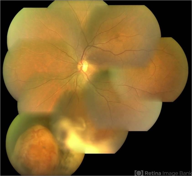
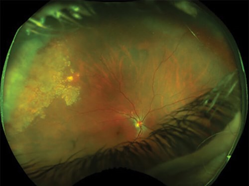
Visual acuity measured 20/40 OS. Fundus examination revealed an elevated, reddish-pink mass located inferotemporally with surrounding hard exudates, subretinal fluid, and mild vitreous cells.
Peripheral retinal telangiectasia and cystoid macular edema were noted.
Optical coherence tomography (OCT) confirmed intraretinal edema with subfoveal fluid, and B-scan ultrasonography demonstrated an acoustically solid, moderately reflective mass measuring 3.5 mm in thickness.
Fluorescein angiography showed early hyperfluorescence with late leakage characteristic of a vascular peripheral tumor.
MRI ruled out systemic malignancy. A diagnosis of idiopathic retinal vasoproliferative tumor (VPT) was made.
The patient underwent cryotherapy followed by intravitreal anti-VEGF therapy, resulting in partial regression and significant improvement in macular anatomy.
Visual acuity stabilized at 20/30, and she remained under careful follow-up.
Disease Entity
Retinal Vasoproliferative Tumor (VPT) is a rare, benign but potentially vision-threatening peripheral retinal vascular mass composed of mixed glial and vascular proliferation.
Historically confused with retinal capillary hemangiomas, VPT is now recognized as a separate entity characterized by exudation, traction, and macular complications.
VPT may be primary (idiopathic) or secondary to pre-existing ocular pathology.
Although benign, the tumor can cause profound retinal damage if untreated due to leakage, chronic macular edema, epiretinal membrane formation, and tractional changes.
Fundus Explorer Pro
Photograph the retinal findings described here with the phone already in your pocket — 22 D optics and built-in illumination in one handheld unit.
From Choroida — the team behind this sitePathophysiology
The exact mechanism of VPT development is uncertain, but several pathological processes contribute:
-
Dominant glial proliferation, mainly astrocytes, forms the bulk of the lesion
-
Reactive vascular proliferation triggered by chronic retinal irritation, inflammation, or ischemia
-
VEGF-driven leakage, leading to exudation and macular involvement
-
Müller cell dysfunction, contributing to retinal thickening
-
Secondary VPTs arising in settings such as:
-
Retinitis pigmentosa
-
Retinal detachment surgery
-
Chronic uveitis
-
Coats disease
-
Retinal dystrophies
-
The interplay of glial and vascular components creates a solid, exudative mass that can induce secondary retinal structural alterations.
Epidemiology
Although rare, certain epidemiologic trends are consistent:
-
Occurs most often in adults aged 20–60 years
-
Slight female predominance in some studies
-
Unilateral in ~75% of cases
-
About 70–80% are idiopathic, with the remainder being secondary
-
Preferentially located inferotemporally
Because symptoms often arise only after macular involvement, many tumors are detected late.
Clinical Features
Symptoms
Patients may present with:
-
Painless visual decline
-
Floaters
-
Photopsia
-
Peripheral field scotoma
-
Distortion (if macula involved)
Signs
Fundus findings typically show:
-
A pink, yellowish, or reddish peripheral mass
-
Size usually 2–4 mm in thickness
-
Hard exudates
-
Subretinal and intraretinal fluid
-
Cystoid macular edema
-
Retinal telangiectasia
-
Vitreous cells
-
Secondary epiretinal membrane
-
Localized traction or retinoschisis
Macular involvement explains most cases of visual loss.
Imaging Findings
OCT
-
Intraretinal cysts
-
Subretinal fluid extending toward the macula
-
ERM formation
-
Outer retinal disruption in chronic cases
FA
-
Early hyperfluorescence
-
Progressive late leakage
-
Prominent feeder and draining vessels
ICGA
-
It better outlines the vascular supply
-
Helps distinguish VPT from choroidal tumors
B-Scan Ultrasound
-
Solid lesion
-
Moderate internal reflectivity
-
No calcifications (helps exclude retinoblastoma)
MRI
-
Mild post-contrast enhancement
-
Differentiates from malignant lesions such as melanoma
Differential Diagnosis
Important entities to differentiate include:
-
Retinal capillary hemangioma (especially in VHL disease)
-
Coats disease
-
Retinoblastoma (in pediatric cases)
-
Peripheral exudative hemorrhagic chorioretinopathy (PEHCR)
-
Choroidal melanoma
-
Reactive gliosis
Accurate differentiation prevents unnecessary aggressive or oncologic treatments.
Diagnosis
Diagnosis is clinical but confirmed by multimodal imaging:
-
Fundus examination
-
OCT for macular assessment
-
FA/ICGA for vascular characterization
-
B-scan ultrasonography for structural evaluation
-
Systemic evaluation for underlying conditions (if secondary VPT suspected)
In unusual cases, referral to an ocular oncology specialist is recommended.
Management
Treatment depends on symptoms, tumor size, and complications.
Observation
For small, non-exudative, asymptomatic tumors.
Cryotherapy
-
Most common treatment
-
Effective for exudative or enlarging lesions
-
Induces regression and reduces leakage
-
May require multiple sessions
Laser Photocoagulation
Useful for small or superficial lesions but less effective alone.
Anti-VEGF Therapy
-
Controls exudation and macular edema
-
Often used adjunctively with cryotherapy
-
Particularly beneficial for VEGF-driven leakage
Steroids
Considered in inflammation-associated secondary VPTs.
Plaque Brachytherapy
Option for large or refractory lesions.
Pars Plana Vitrectomy
For:
-
Tractional complications
-
Dense vitreous hemorrhage
-
ERM removal
-
Retinal detachment
Prognosis
Prognosis varies depending on:
-
Tumor size
-
Degree of macular involvement
-
Timeliness of treatment
Early intervention typically stabilizes or improves vision, while chronic macular changes (outer retinal atrophy, fibrosis) limit visual recovery. Secondary VPTs carry a risk of recurrence if the underlying disease is uncontrolled.
Prevention
No direct prevention for idiopathic VPT exists. However:
-
Early treatment of ocular inflammatory or ischemic diseases
-
Regular retinal follow-up in high-risk patients
-
Prompt evaluation of unexplained exudation or macular edema


Document what you see
Two smartphone imaging tools built for everyday clinic use — one for the slit lamp, one for the fundus.
From Choroida — the team behind this siteReferences
-
Shields CL, Shields JA. Vasoproliferative tumors of the retina. Retina.
-
Heimann H, et al. Management of retinal vasoproliferative tumors. Ophthalmology.
-
Gass JD. Peripheral retinal tumors and their differential diagnosis.
-
Venkatesh P, et al. Anti-VEGF therapy in retinal vasoproliferative tumors.
-
Singh AD, et al. Imaging and management strategies for peripheral retinal tumors.
Test yourself
A few questions straight from this article.
-
Retinal vasoproliferative tumor is best described as which kind of peripheral retinal lesion?
The article defines VPT as a rare, benign but potentially vision-threatening peripheral retinal vascular mass made up of mixed glial and vascular proliferation. -
Which cell type forms the bulk of the lesion in retinal vasoproliferative tumor?
Dominant glial proliferation, mainly astrocytes, makes up most of the mass, with reactive vascular proliferation layered on top of it. -
Roughly what proportion of retinal vasoproliferative tumors are idiopathic rather than secondary?
About 70–80% of VPTs are primary or idiopathic; the remainder arise secondary to conditions such as retinitis pigmentosa, chronic uveitis or Coats disease. -
Where is a retinal vasoproliferative tumor most often located?
VPTs show a clear preference for the inferotemporal peripheral retina, which is where the case lesion was also found. -
What pattern does fluorescein angiography show in retinal vasoproliferative tumor?
FA shows early hyperfluorescence followed by progressive late leakage, together with prominent feeder and draining vessels. -
Which B-scan feature of a retinal vasoproliferative tumor helps exclude retinoblastoma?
On ultrasound a VPT is solid with moderate internal reflectivity and contains no calcification, which argues against retinoblastoma. -
Retinal vasoproliferative tumor was historically confused with which other retinal vascular tumor?
VPT was long grouped with retinal capillary haemangioma but is now recognised as a separate entity defined by exudation, traction and macular complications. -
Which treatment is most commonly used for an exudative or enlarging retinal vasoproliferative tumor?
Cryotherapy is the most common treatment for exudative or enlarging lesions, inducing regression and reducing leakage, though it may need several sessions. -
What is the main role of intravitreal anti-VEGF therapy in retinal vasoproliferative tumor?
Anti-VEGF targets the VEGF-driven leakage behind the exudation and macular oedema, and is usually given alongside cryotherapy rather than alone. -
What raises the risk of recurrence in a secondary retinal vasoproliferative tumor?
Secondary VPTs recur when the underlying ocular condition that drove them, such as chronic uveitis or a retinal dystrophy, remains uncontrolled.
