
Case Study
A 29-year-old woman with a 3-year history of systemic lupus erythematosus (SLE) presented with sudden-onset blurry vision in her left eye, accompanied by mild ocular discomfort and increased photosensitivity.
She had been non-compliant with her hydroxychloroquine treatment for the past two months due to gastrointestinal side effects.
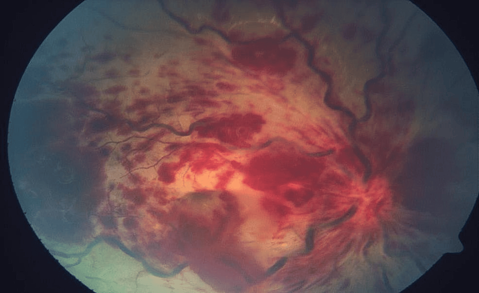
Ophthalmic examination revealed cotton-wool spots, microaneurysms, and scattered intraretinal hemorrhages along the posterior pole, with perivascular sheathing more pronounced in the superior arcade.
OCT demonstrated inner retinal ischemia with paracentral acute middle maculopathy-like changes. Fluorescein angiography confirmed areas of capillary non-perfusion, vascular leakage, and delayed arteriovenous transit time.
Laboratory testing showed elevated anti-dsDNA, low complement levels, and increased ESR, indicating active systemic disease.
She was immediately started on high-dose systemic corticosteroids and consulted rheumatology for immunosuppressive escalation.
Despite treatment, the affected eye recovered only to 20/60 due to persistent macular ischemia, highlighting the aggressive nature of SLE-associated retinal vasculopathy.
Disease Entity
Systemic lupus erythematosus (SLE) is a chronic autoimmune disorder affecting multiple organ systems through immune complex deposition and complement-mediated inflammation.
Ocular involvement occurs in 20–34% of patients and can affect virtually any structure of the eye, from the adnexa to the retina and optic nerve.
Retinal and choroidal manifestations are among the most vision-threatening complications, often correlating strongly with systemic disease activity.
Ocular complications range from mild keratoconjunctivitis sicca to severe retinal vaso-occlusive disease and lupus choroidopathy, making ophthalmologists integral to early detection of systemic flares and guiding timely treatment.
Fundus Explorer Pro
Photograph the retinal findings described here with the phone already in your pocket — 22 D optics and built-in illumination in one handheld unit.
From Choroida — the team behind this sitePathophysiology
The main pathogenic mechanism in SLE ocular disease is immune complex deposition within small vessels of the eye, leading to:
-
Complement activation and inflammation
-
Endothelial damage and capillary occlusion
-
Vascular leakage and ischemia
-
Breakdown of the blood–retinal barrier
The retina is particularly vulnerable due to its dense microvascular network.
Other mechanisms include:
-
Autoantibody-mediated vascular injury
-
Procoagulant state leading to arterial or venous occlusion
-
Choroidal ischemia secondary to complement activation
-
Drug-induced toxicity (notably hydroxychloroquine)
Epidemiology
-
Ocular involvement occurs in up to one-third of SLE patients.
-
Retinal involvement is reported in 3–15%, typically during active systemic flares.
-
Severe vaso-occlusive retinopathy is more common in patients with antiphospholipid syndrome (APS) or high disease activity.
-
Lupus choroidopathy is strongly associated with renal involvement and hypertension.
Clinical Features
Ocular complications of SLE involve both anterior and posterior segment structures.
1. Anterior Segment Manifestations:
-
Keratoconjunctivitis sicca (most common)
-
Episcleritis or scleritis
-
Peripheral ulcerative keratitis
These are usually associated with systemic disease activity.
2. Retinal Manifestations (Most Vision-Threatening):
Lupus Retinopathy:
-
Cotton-wool spots
-
Intraretinal hemorrhages
-
Microaneurysms
-
Retinal vascular leakage
-
Venous dilation and tortuosity
-
Perivascular sheathing (“vasculitis”)
Severe Vaso-Occlusive Lupus Retinopathy:
-
Branch or central retinal vein occlusion
-
Arterial occlusions
-
Purtscher-like retinopathy
-
Wide areas of capillary non-perfusion
-
Acute macular ischemia
-
Neovascularization and vitreous hemorrhage in late stages
3. Choroidal Manifestations:
-
Lupus choroidopathy
-
Serous retinal detachments due to choroidal ischemia
-
RPE folds and mottling
4. Optic Nerve Involvement:
-
Optic neuritis
-
Ischemic optic neuropathy
-
Papilledema associated with CNS involvement
Examination Findings
-
OCT: inner retinal thinning, ischemic changes, serous retinal detachment in choroidopathy.
-
FA: delayed filling, leakage, non-perfusion, arterial attenuation.
-
ICG angiography: choroidal hypoperfusion and multifocal ischemic spots.
-
Visual fields: arcuate or paracentral defects depending on ischemia.

Differential Diagnosis
-
Diabetic retinopathy
-
Hypertensive retinopathy
-
Infectious retinal vasculitis (TB, syphilis, toxoplasmosis)
-
Retinal vasculitis of other autoimmune diseases
-
Antiphospholipid syndrome retinopathy
-
Hydroxychloroquine retinal toxicity (distinct pattern)
Diagnosis
Diagnosis is based on clinical examination and diagnostic imaging combined with systemic assessments.
Essential Investigations:
-
Fluorescein angiography (FA)
-
OCT and OCT-A
-
ICG for choroidal involvement
-
Autoimmune panel: ANA, dsDNA, SSA/SSB
-
Complement levels (C3, C4)
-
APS testing (anticardiolipin, lupus anticoagulant, β2 glycoprotein)
Management
Treatment targets both ocular inflammation and systemic disease.
1. Systemic Immunosuppression
Mainstay of treatment.
-
High-dose corticosteroids (IV methylprednisolone in severe disease)
-
Immunosuppressants:
-
Mycophenolate mofetil
-
Azathioprine
-
Cyclophosphamide (severe vasculopathy or choroidopathy)
-
Methotrexate
-
2. Biologic Therapy
Used in refractory disease:
-
Belimumab
-
Rituximab (especially in vaso-occlusive retinopathy)
3. Anticoagulation
Required if APS is present.
-
Warfarin or DOACs
-
Aspirin may be used adjunctively
4. Ocular-Specific Management
-
Panretinal photocoagulation for ischemia-induced neovascularization
-
Anti-VEGF injections for macular edema
-
Surgical intervention for complications such as vitreous hemorrhage or tractional detachment
5. Management of Dry Eye
-
Artificial tears
-
Topical cyclosporine or lifitegrast
Prognosis
-
Mild lupus retinopathy often improves with systemic control.
-
Severe vaso-occlusive disease carries a poor prognosis, especially with macular ischemia.
-
Lupus choroidopathy prognosis improves with rapid treatment but may be associated with progression of systemic renal disease.
-
Early diagnosis and aggressive systemic therapy are critical in preventing permanent vision loss.
Prevention & Follow-Up
-
Regular ophthalmic evaluation for all SLE patients
-
More frequent monitoring during disease flares
-
Screening for hydroxychloroquine toxicity
-
Close communication with rheumatology
-
Prompt evaluation of any new visual symptoms


Document what you see
Two smartphone imaging tools built for everyday clinic use — one for the slit lamp, one for the fundus.
From Choroida — the team behind this siteReferences
-
Jabs DA. Ocular manifestations of systemic lupus erythematosus. Curr Opin Ophthalmol.
-
Rangaswamy R, et al. Retinal vascular involvement in SLE. Lupus Science & Medicine.
-
Nguyen QD, et al. Lupus choroidopathy and vaso-occlusive retinopathy. Ophthalmology.
-
Arevalo JF, et al. Severe retinal vaso-occlusive disease in SLE: Diagnosis and management. Retina Journal.
-
Yeh S. Autoimmune retinopathies and systemic autoimmune disease.
Test yourself
A few questions straight from this article.
-
In what proportion of patients with systemic lupus erythematosus does ocular involvement occur?
Ocular involvement affects roughly one-third of SLE patients and can involve any ocular structure, from the adnexa to the retina and optic nerve. -
What is the principal pathogenic mechanism of ocular disease in systemic lupus erythematosus?
Immune complexes deposited in small ocular vessels drive complement activation, endothelial damage, capillary occlusion and breakdown of the blood-retinal barrier. -
How often is retinal involvement reported in systemic lupus erythematosus?
Retinal involvement is reported in 3-15% of SLE patients and typically appears during active systemic flares. -
Which anterior segment finding is the most common ocular manifestation of systemic lupus erythematosus?
Keratoconjunctivitis sicca is the commonest anterior segment manifestation in SLE and is usually managed with artificial tears, topical cyclosporine or lifitegrast. -
Lupus choroidopathy shows a strong association with which systemic features?
Lupus choroidopathy is strongly linked to renal disease and hypertension, and its course may parallel progression of systemic renal disease. -
Severe vaso-occlusive lupus retinopathy is most likely in patients who also have which condition?
Severe vaso-occlusive retinopathy is more frequent in patients with antiphospholipid syndrome or high systemic disease activity, and anticoagulation is required when APS is present. -
What does indocyanine green angiography typically demonstrate in lupus choroidopathy?
ICG angiography in lupus choroidopathy reveals choroidal hypoperfusion and multifocal ischemic spots, while OCT may show serous retinal detachment. -
What is the mainstay of treatment for sight-threatening ocular complications of systemic lupus erythematosus?
Systemic immunosuppression is the mainstay, using high-dose corticosteroids (IV methylprednisolone in severe disease) with agents such as mycophenolate, azathioprine or cyclophosphamide. -
Which biologic agent is highlighted for refractory vaso-occlusive lupus retinopathy?
Biologics are reserved for refractory disease, with rituximab specifically favoured in vaso-occlusive retinopathy and belimumab as the other named option. -
Which form of lupus eye disease carries the poorest visual prognosis?
Mild lupus retinopathy often improves once systemic disease is controlled, whereas severe vaso-occlusive disease with macular ischemia carries a poor prognosis.
