
Case Study
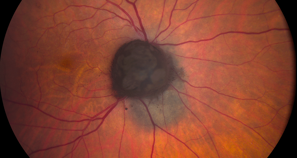
A 56-year-old woman presented with a pigmented lesion in the posterior pole of her left eye, discovered incidentally during a routine ophthalmic examination.
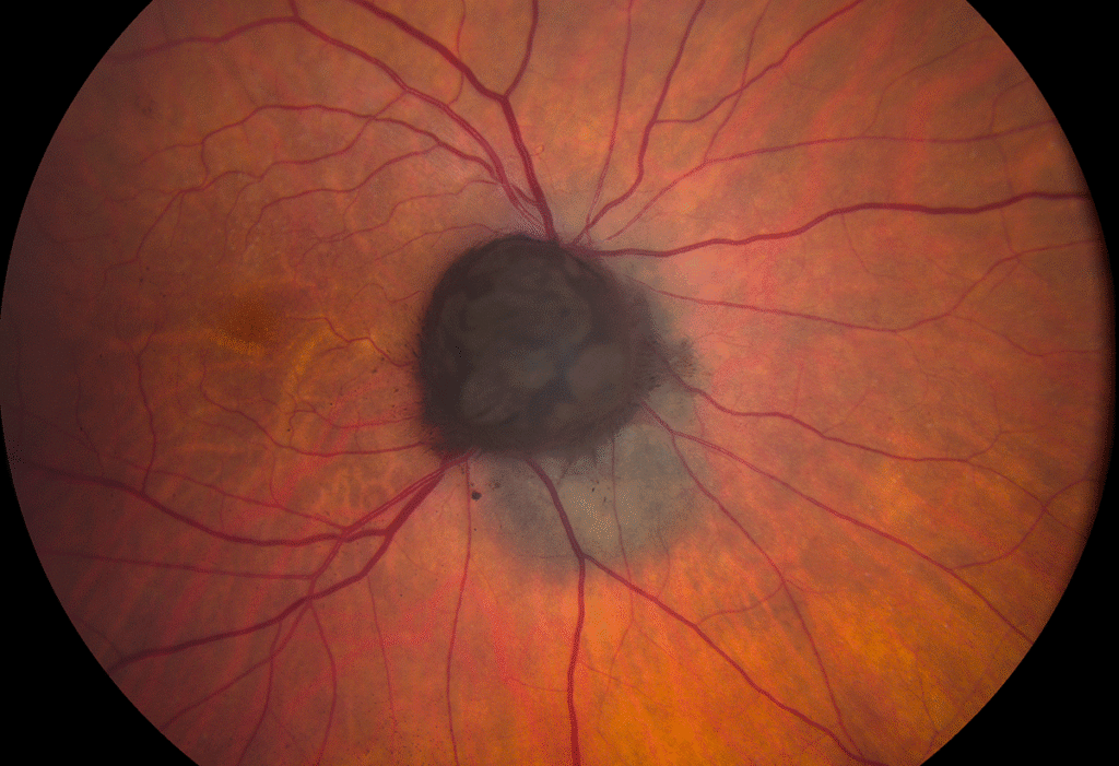
She had no visual complaints, and her best-corrected visual acuity was 20/20 in both eyes. Fundus examination revealed a dark brown, dome-shaped lesion at the peripapillary region with well-defined margins.
Optical coherence tomography (OCT) demonstrated a mildly elevated lesion with intact overlying retina, while B-scan ultrasonography showed medium internal reflectivity.
Given the lesion’s appearance, a differential diagnosis including choroidal melanoma was considered.
However, long-term observation revealed no growth, confirming the diagnosis of Choroidal Melanocytoma—a benign but deceptively pigmented tumor.
Disease Entity
Choroidal Melanocytoma is a rare, benign pigmented tumor of the uveal tract, typically composed of deeply pigmented melanocytes.
Although histologically benign, its clinical presentation can mimic malignant melanoma, making accurate differentiation essential to avoid unnecessary enucleation.
It most commonly affects the optic disc or peripapillary region, but can occasionally arise in the choroid or ciliary body.
Fundus Explorer Pro
Photograph the retinal findings described here with the phone already in your pocket — 22 D optics and built-in illumination in one handheld unit.
From Choroida — the team behind this sitePathophysiology
Choroidal melanocytomas arise from melanocytes—neural crest-derived cells responsible for ocular pigmentation.
These lesions consist of round or polygonal cells containing abundant melanin granules that can obscure nuclear details.
Despite being benign, they may undergo necrosis or cause secondary inflammation, leading to local complications such as vitreous seeding or retinal edema.
Rarely, transformation into malignant melanoma has been reported, though this remains exceedingly uncommon.
Epidemiology
Choroidal melanocytoma is most frequently diagnosed in middle-aged adults, with a slight predilection for females. There appears to be no racial predilection, although darker-skinned individuals may have a higher incidence of ocular melanocytic lesions in general.
The optic disc is the most common site of involvement, followed by the peripapillary choroid.
Clinical Features
Patients are often asymptomatic, and the lesion is usually discovered incidentally.
When symptomatic, visual disturbance may result from compression of the optic nerve, secondary serous retinal detachment, or vitreous seeding.
Typical clinical findings include:
-
A dark brown to black, elevated mass on the optic disc or adjacent choroid
-
Sharp, well-defined margins
-
Occasional orange pigmentation or mottling due to necrosis
-
Overlying retinal edema or subretinal fluid in symptomatic cases
Examination Findings
-
Fundus Examination: Deeply pigmented, dome-shaped lesion with sharp borders and occasional overlying exudation.
-
OCT: Localized elevation with intact retinal layers, sometimes showing subretinal fluid.
-
B-scan Ultrasonography: Medium internal reflectivity (distinguishing it from the low reflectivity of melanoma).
-
Fluorescein Angiography: Hypofluorescence throughout, as dense melanin blocks fluorescence.
-
Autofluorescence Imaging: Hypoautofluorescent due to heavy pigmentation.
Differential Diagnosis
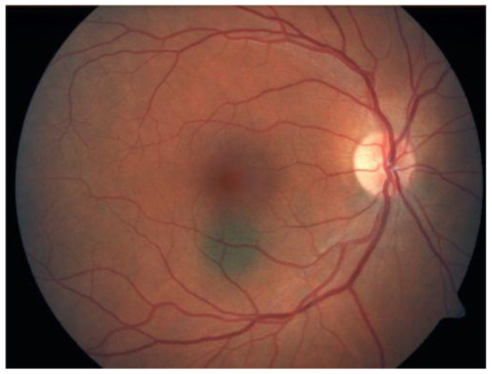
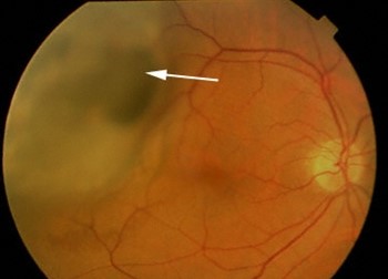
Because of its pigmentation and elevation, Choroidal Melanocytoma can easily be mistaken for:
-
Choroidal melanoma – tends to show orange pigment, lipofuscin, and low internal reflectivity
-
Choroidal nevus – typically flat or slightly elevated with less dense pigmentation
-
Adenoma of the retinal pigment epithelium (RPE) – may have irregular margins and less pigmentation
-
Optic disc melanoma or metastasis – often accompanied by rapid growth and visual symptoms
Diagnosis
Diagnosis is based primarily on characteristic clinical and imaging findings. Stability over time is the key diagnostic indicator confirming benignity.
-
Observation: Most lesions are monitored with periodic fundus photography and OCT to document stability.
-
Fine-needle aspiration biopsy (FNAB): Rarely indicated, reserved for cases where malignancy cannot be excluded through imaging alone.
Management
Observation is the mainstay of management for choroidal melanocytoma. Regular follow-up every 6–12 months is recommended to detect any signs of growth or complications.
-
Indications for intervention:
-
Documented lesion enlargement
-
Secondary complications such as vitreous seeding or retinal detachment
-
Diagnostic uncertainty
-
-
Treatment options:
-
Laser photocoagulation or radiotherapy in selected cases with exudative complications
-
Enucleation only if malignant transformation or extensive necrosis is confirmed
-
Prognosis
The prognosis for choroidal melanocytoma is excellent in most cases, as these tumors remain stable for years or decades.
However, patients should be educated about the importance of follow-up, since complications such as necrosis, inflammation, or rare malignant transformation can alter outcomes.
Vision may be affected if the lesion involves the optic disc or causes secondary retinal damage.
Prevention
There are no established preventive measures, as choroidal melanocytoma is a sporadic condition without known modifiable risk factors.
However, early detection and regular ophthalmic evaluations are crucial for differentiation from more aggressive lesions.


Document what you see
Two smartphone imaging tools built for everyday clinic use — one for the slit lamp, one for the fundus.
From Choroida — the team behind this siteReferences
-
Shields JA, Shields CL. Intraocular Tumors: An Atlas and Textbook, 3rd Edition. Lippincott Williams & Wilkins; 2016.
-
Arepalli S, Kaliki S, Shields CL. Choroidal melanocytoma: Clinical features and natural course in 115 cases. Br J Ophthalmol. 2015;99(3):377–382.
-
Singh AD, Damato BE, Pe’er J, et al. Clinical Ophthalmic Oncology: Uveal Tumors. Springer; 2019.
-
Demirci H, Shields CL, Eagle RC Jr. Choroidal melanocytoma: Clinicopathologic correlation in 17 cases. Arch Ophthalmol. 2001;119(12):1767–1774.
Test yourself
A few questions straight from this article.
-
How is choroidal melanocytoma best characterised?
Choroidal melanocytoma is a rare benign tumour made of deeply pigmented melanocytes; recognising its benign nature avoids unnecessary enucleation. -
Which site is most commonly involved by choroidal melanocytoma?
The optic disc is the commonest location, followed by the peripapillary choroid; ciliary body involvement is unusual. -
From which cells does choroidal melanocytoma arise?
The tumour is composed of round or polygonal melanocytes of neural crest origin, packed with melanin granules that obscure nuclear detail. -
What does B-scan ultrasonography typically show in choroidal melanocytoma?
Medium internal reflectivity on B-scan helps separate melanocytoma from choroidal melanoma, which characteristically shows low internal reflectivity. -
What is seen on fluorescein angiography in choroidal melanocytoma?
Dense melanin blocks fluorescence, so the lesion stays hypofluorescent; it is likewise hypoautofluorescent on autofluorescence imaging. -
Which feature favours choroidal melanoma rather than choroidal melanocytoma?
Choroidal melanoma tends to display orange lipofuscin pigment and low internal reflectivity, while melanocytoma shows sharp margins and medium reflectivity. -
Which finding confirms the benign nature of a choroidal melanocytoma?
Stability over time is the key diagnostic indicator, documented with serial fundus photography and OCT rather than routine biopsy. -
What is the mainstay of management for choroidal melanocytoma?
Observation is the mainstay, with review every 6 to 12 months to detect growth or complications such as vitreous seeding. -
When is enucleation justified in a patient with choroidal melanocytoma?
Enucleation is reserved for confirmed malignant transformation or extensive necrosis; exudative complications may instead be treated with laser or radiotherapy. -
What is the usual long-term course of choroidal melanocytoma?
Prognosis is excellent because these tumours usually stay stable for years or decades, though follow-up matters since necrosis, inflammation and rare malignant change can occur.
