
Case Study
A 58-year-old man with a history of nasopharyngeal carcinoma presented with sudden, painless vision loss in his right eye, occurring six months after completing radiotherapy.
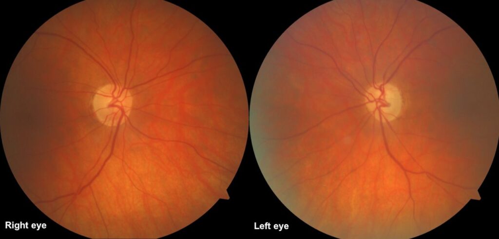
Visual acuity had dropped from 6/6 to counting fingers within a few days. Fundus examination revealed optic disc pallor with blurred margins.
Optical coherence tomography (OCT) demonstrated thinning of the retinal nerve fiber layer, and MRI excluded tumor recurrence.
Based on clinical history and findings, a diagnosis of Radiation-Induced Optic Neuropathy (RION) was made.
Disease Entity
Radiation-Induced Optic Neuropathy (RION) is a rare but devastating delayed complication of radiotherapy involving structures near the optic nerve, chiasm, or orbit.
It is characterized by sudden, painless, and often irreversible vision loss, typically occurring months to years after radiation exposure.
The condition results from vascular and axonal damage within the optic nerve due to radiation-induced ischemia and necrosis.
Fundus Explorer Pro
Photograph the retinal findings described here with the phone already in your pocket — 22 D optics and built-in illumination in one handheld unit.
From Choroida — the team behind this sitePathophysiology
RION develops when high-dose ionizing radiation injures the small vessels supplying the optic nerve. This leads to occlusive vasculopathy, ischemia, demyelination, and axonal loss.
Histopathological findings reveal hyalinized vessels, demyelination, and gliosis. The risk increases when the cumulative radiation dose exceeds 50 Gy to the optic apparatus.
Concomitant chemotherapy, diabetes, and hypertension may exacerbate the damage. Unlike compressive or infiltrative optic neuropathies, RION is primarily ischemic in origin.
The latency period can vary from a few months to several years, reflecting the slow progression of vascular compromise and delayed cellular death within the optic nerve tissue.
Epidemiology
The incidence of RION is estimated to be between 0.5% and 3% in patients receiving cranial or orbital irradiation.
It typically occurs 6 to 36 months after therapy, with no gender predilection. Tumors most commonly associated include nasopharyngeal carcinoma, pituitary adenoma, meningioma, and sinonasal malignancies. Advanced radiotherapy techniques such as stereotactic radiosurgery, proton beam therapy, and intensity-modulated radiotherapy (IMRT) have reduced but not eliminated the risk.
Clinical Features
Patients usually report sudden, painless vision loss, often unilateral but occasionally bilateral when both optic nerves are within the radiation field. Visual field defects, such as central or cecocentral scotomas, are common. Some patients may retain partial peripheral vision.
On examination, the optic disc may initially appear normal but later becomes swollen and pale. With time, optic atrophy develops. Pain, inflammation, or systemic symptoms are typically absent, differentiating RION from optic neuritis or tumor recurrence.
Examination Findings (Summary):
-
Visual acuity: Severe decrease, often to counting fingers or worse
-
Pupils: Relative afferent pupillary defect (RAPD) in unilateral cases
-
Fundus: Optic disc edema progressing to pallor
-
Visual field: Central or altitudinal scotoma
-
OCT: RNFL thinning
-
MRI: Optic nerve enhancement without mass effect
Differential Diagnosis
RION can resemble several other causes of optic neuropathy. Key differentials include:
-
Optic neuritis (inflammatory demyelination)
-
Compressive optic neuropathy (tumor recurrence or metastasis)
-
Ischemic optic neuropathy (non-arteritic or arteritic)
-
Infectious optic neuropathy (toxoplasmosis, tuberculosis)
-
Infiltrative or toxic optic neuropathies
Diagnostic Approach
Diagnosis relies on clinical history of prior radiation exposure and exclusion of other causes. MRI with contrast is crucial—it typically shows enhancement and swelling of the affected optic nerve without evidence of tumor recurrence. OCT helps monitor structural changes and exclude other retinal pathologies.
Visual evoked potentials (VEP) may demonstrate reduced amplitude and delayed latency, consistent with axonal loss. Fluorescein angiography may show disc leakage in early stages.
Management
Unfortunately, there is no proven effective therapy for RION. Treatment focuses on early recognition and supportive care.
Management Strategies:
-
High-dose corticosteroids: Often used empirically in acute phases, though benefits are inconsistent.
-
Hyperbaric oxygen therapy (HBOT): May improve oxygenation and delay progression if started early.
-
Bevacizumab (anti-VEGF): Used experimentally in some cases to reduce vascular permeability.
-
Neuroprotective agents: Limited evidence, but some clinicians use antioxidants or vitamins.
-
Visual rehabilitation: Essential for patients with permanent vision loss.
Preventive measures remain the most effective strategy—limiting radiation dose to the optic nerve and chiasm, careful treatment planning, and avoiding overlapping radiation fields.

Prognosis
The prognosis of RION is generally poor, with most patients experiencing irreversible vision loss. Rarely, partial recovery occurs if intervention begins very early. Bilateral involvement predicts worse outcomes. Long-term follow-up and counseling are vital for patient adaptation to visual disability.
Prevention
Prevention is crucial, as established damage cannot be reversed. Radiation oncologists must ensure that the radiation dose to the optic apparatus stays below 50 Gy, using advanced radiotherapy planning systems to spare visual structures. Monitoring patients for early visual changes and prompt referral to ophthalmology can mitigate severe outcomes.


Document what you see
Two smartphone imaging tools built for everyday clinic use — one for the slit lamp, one for the fundus.
From Choroida — the team behind this siteReferences
-
Miller NR, et al. Radiation-Induced Optic Neuropathy: Clinical and Pathophysiologic Review. J Neuroophthalmol.
-
Guy J, et al. Radiation Optic Neuropathy: A Review of Pathogenesis and Management. Surv Ophthalmol.
-
Danesh-Meyer HV. Radiation-Induced Optic Neuropathy: Current Understanding. Eye (Lond).
-
Parsons JT, et al. Dose-Response Relationships for Radiation-Induced Optic Neuropathy. Int J Radiat Oncol Biol Phys.
-
Levin LA. Ischemic and Radiation-Induced Optic Neuropathies: Mechanisms and Therapy. Prog Retin Eye Res.
-
Mishra KK, et al. Risk Factors and Clinical Course of Radiation Optic Neuropathy. Pract Radiat Oncol.
Test yourself
A few questions straight from this article.
-
Which description best characterises radiation-induced optic neuropathy (RION)?
RION is a delayed complication of radiotherapy near the optic nerve, chiasm or orbit, producing sudden painless and usually irreversible visual loss months to years after exposure. -
What is the primary mechanism of optic nerve injury in radiation-induced optic neuropathy?
Ionising radiation injures the small vessels supplying the optic nerve, producing occlusive vasculopathy with ischaemia, demyelination and axonal loss. RION is therefore ischaemic rather than compressive or inflammatory. -
Above which cumulative radiation dose to the optic apparatus does the risk of RION rise sharply?
Risk increases once the cumulative dose to the optic apparatus exceeds 50 Gy, which is why radiotherapy planning aims to keep the optic nerve and chiasm below this threshold. -
What is the estimated incidence of RION in patients receiving cranial or orbital irradiation?
RION is rare, occurring in roughly 0.5% to 3% of irradiated patients, usually 6 to 36 months after therapy and with no gender predilection. -
Which malignancy is classically associated with radiation-induced optic neuropathy?
Tumours treated with fields close to the optic apparatus carry the risk, most commonly nasopharyngeal carcinoma, pituitary adenoma, meningioma and sinonasal malignancies. -
Which MRI appearance supports a diagnosis of radiation-induced optic neuropathy?
Contrast MRI typically shows enhancement and swelling of the affected optic nerve with no evidence of tumour recurrence, which is what makes it central to the diagnosis. -
Which feature best separates radiation-induced optic neuropathy from optic neuritis?
Pain, inflammation and systemic symptoms are typically absent in RION, which is what differentiates it from optic neuritis and from tumour recurrence. -
Which statement reflects the current treatment position for radiation-induced optic neuropathy?
There is no proven effective therapy for RION. Management centres on early recognition, supportive care and visual rehabilitation, with prevention being the most effective strategy. -
Which therapy may improve oxygenation and delay progression of RION if started early?
Hyperbaric oxygen therapy may improve oxygenation of the ischaemic nerve and delay progression when begun early, alongside empirical corticosteroids and experimental bevacizumab. -
What is the usual visual prognosis of radiation-induced optic neuropathy?
Most patients suffer irreversible vision loss; only rarely does partial recovery follow very early intervention, and bilateral involvement predicts a worse outcome.
