
Disease entity
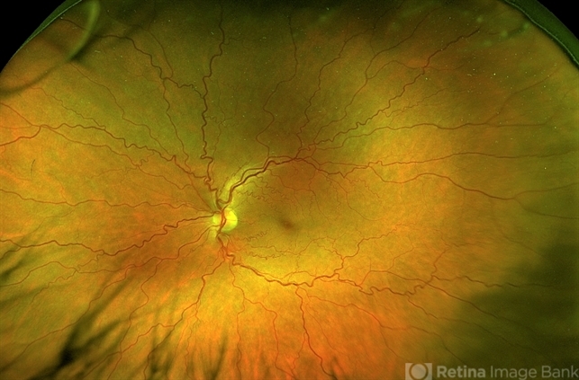
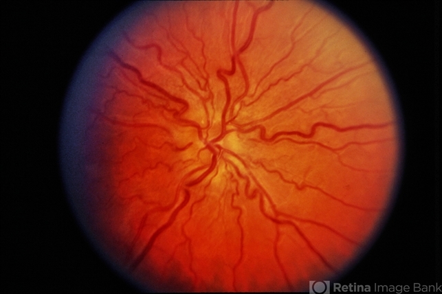
Retinal vascular tortuosity is characterized by an abnormal twisting or coiling of the retinal blood vessels, often observed in both arteries and veins.

While retinal vascular tortuosity can sometimes be an incidental finding, it is also associated with various systemic conditions, such as vascular and connective tissue disorders, congenital heart diseases, and certain genetic syndromes.
Retinal tortuosity can be present at birth or develop over time and may signal underlying vascular fragility or systemic pathology.
This condition’s etiology, clinical significance, and relationship to systemic disease make it a relevant finding in both pediatric and adult patients.
Pathophysiology
The mechanism behind vascular tortuosity involves structural abnormalities in the blood vessel wall. When the balance between blood flow and vessel elasticity is disrupted, vessels may adopt a tortuous path.
Vascular tortuosity often reflects weakened vessel walls, commonly seen in connective tissue disorders such as Ehlers-Danlos syndrome (EDS) or Loeys-Dietz syndrome.
Increased tortuosity can cause variations in blood flow and pressure, increasing the risk of vascular complications such as retinal vein occlusion or hemorrhage.
The primary mechanisms involved in tortuosity are thought to include genetic mutations affecting collagen and elastin in the vessel walls, endothelial dysfunction, and systemic blood pressure irregularities.
Fundus Explorer Pro
Photograph the retinal findings described here with the phone already in your pocket — 22 D optics and built-in illumination in one handheld unit.
From Choroida — the team behind this siteEpidemiology
Retinal vascular tortuosity has a variable prevalence, given its association with numerous systemic conditions.
It is observed across all age groups and can be hereditary or sporadic. In some cases, it occurs as an isolated retinal finding, while in others, it is part of a syndrome such as Marfan syndrome or systemic hypertension.
The prevalence of retinal tortuosity is higher in individuals with connective tissue disorders and certain familial vascular syndromes.
Clinical Features
Patients with retinal vascular tortuosity may be asymptomatic or may present with a variety of visual complaints:
- Visual Blurring: Temporary blurring, often related to changes in blood flow.
- Photophobia: Increased sensitivity to light.
- Floaters: If hemorrhage or exudates are present, floaters may occur.
- Hemorrhagic Complications: In severe cases, spontaneous retinal hemorrhages may occur, particularly in individuals with vascular fragility syndromes.
Retinal vascular tortuosity is typically evident on fundoscopic examination, showing undulating or coiled arteries and veins, often most pronounced at the posterior pole.

Arteries may also appear slightly dilated or narrowed, depending on the underlying condition.
Examination Findings
- Fundoscopic Examination: Tortuosity is easily visualized, especially in the larger vessels at the posterior pole. No other retinal abnormalities may be present, although in cases of severe tortuosity, hemorrhages or cotton-wool spots may appear.
- OCT Angiography: This may be used to assess vessel structure and blood flow, particularly helpful in detecting subclinical vascular changes.
- Fluorescein Angiography (FA): Useful for identifying microvascular anomalies, abnormal leakages, or areas of capillary non-perfusion in severe cases.
Differential Diagnosis
- Retinopathy of prematurity
- Hypertensive retinopathy
- Marfan syndrome
- Ehlers-Danlos syndrome (EDS)
- Familial retinal arterial tortuosity
- Coats disease
Management
Management primarily focuses on monitoring and addressing any underlying systemic conditions. In asymptomatic cases, regular ophthalmic examinations are recommended to detect any progression or complications such as retinal hemorrhage or ischemia.
For patients with syndromic associations like EDS, interdisciplinary management is essential to mitigate the risk of systemic vascular events.
- Observation: Regular follow-up with fundus exams to monitor for retinal hemorrhages or signs of ischemia.
- Protective Measures: Patients with vascular conditions are advised to avoid high-impact activities that could strain blood vessels.
- Systemic Evaluation: Genetic counseling and systemic screening may be indicated, especially if tortuosity is linked to connective tissue disorders.
Prognosis
The prognosis for patients with isolated retinal vascular tortuosity is generally favorable, with most cases remaining stable and not impacting vision significantly.
However, in cases linked with systemic vascular disorders, the prognosis may vary, necessitating lifelong monitoring and systemic management to prevent complications.


Document what you see
Two smartphone imaging tools built for everyday clinic use — one for the slit lamp, one for the fundus.
From Choroida — the team behind this siteReferences
- Manschot, W. A. (1963). Vascular retinal lesions: The incidence and cause of vascular tortuosity of retinal vessels. American Journal of Ophthalmology, 55(4), 538-543.
- Loeys, B. L., & Dietz, H. C. (2008). The loeys-dietz syndrome. Annual Review of Genomics and Human Genetics, 9, 407-428.
- Augsburger, J. J., & Shields, J. A. (1980). Vascular tortuosity of the retinal vessels. American Journal of Ophthalmology, 90(6), 822-824.
- Konieczka, K., & Flammer, J. (2016). Vascular dysregulation in the pathogenesis of retinal diseases. Eye and Brain, 8, 149-155.
