
Case Study
A 60-year-old male presented with complaints of sudden onset blurred vision and floaters in his left eye for the past week.
He had a history of poorly controlled diabetes mellitus and hypertension. There was no history of trauma or previous ocular surgeries.
Examination Results:
- Visual Acuity: 20/20 in the right eye, 20/80 in the left eye.
- Fundus Examination: Extensive areas of retinal whitening and hemorrhages in the mid-peripheral retina of the left eye. Cotton wool spots were observed.
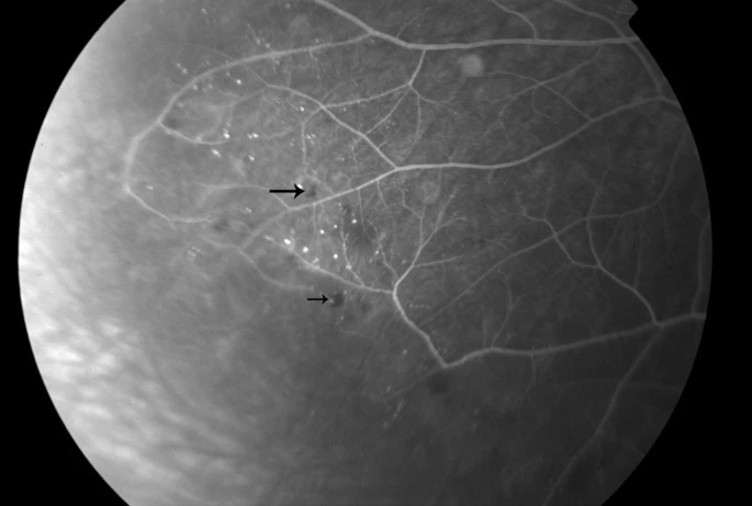
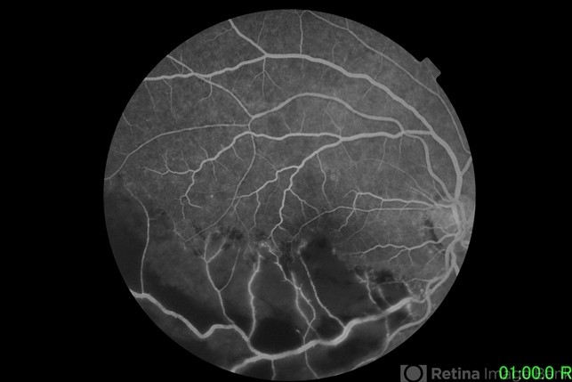
- Fluorescein Angiography: Large areas of capillary nonperfusion and leakage in the affected regions.
- Optical Coherence Tomography (OCT): Significant macular edema and intraretinal fluid in the left eye.
- Systemic Workup: Elevated HbA1c levels and blood pressure of 160/100 mmHg.
The patient was diagnosed with Retinal Capillary Nonperfusion according to previous examination results.
Retinal capillary nonperfusion disease entity
Retinal capillary nonperfusion is a complex and critical disease entity characterized by the inadequate or complete loss of blood flow within the intricate capillary networks of the retina.
It results from a variety of underlying conditions such as diabetic retinopathy, retinal vein occlusions, or retinal ischemia.
This disruption in perfusion can lead to severe visual impairment by compromising the essential retinal structures responsible for vision.
The situation may further worsen through the induction of neovascularization, a process where new, often fragile, and leaky blood vessels form in response to ischemic stress.
This can culminate in significant complications such as macular edema, where fluid accumulates in the central part of the retina, and vitreous hemorrhage, where blood leaks into the gel-like substance filling the eye.
Early diagnosis, vigilant monitoring, and timely intervention are crucial to mitigate the risk of permanent vision loss.
Fundus Explorer Pro
Photograph the retinal findings described here with the phone already in your pocket — 22 D optics and built-in illumination in one handheld unit.
From Choroida — the team behind this siteRetinal capillary nonperfusion Pathophysiology
Retinal capillary nonperfusion occurs when there is an obstruction in the small blood vessels supplying the retina.
This can result from a variety of causes, including diabetic retinopathy, retinal vein occlusion, and hypertensive retinopathy.
The loss of capillary perfusion leads to retinal ischemia, triggering a cascade of pathological changes such as increased vascular permeability, retinal edema, and neovascularization.
Persistent ischemia can cause irreversible damage to the retinal tissue, leading to vision loss.
Retinal capillary nonperfusion Epidemiology
Retinal capillary nonperfusion is commonly associated with systemic diseases such as diabetes mellitus and hypertension.
It predominantly affects older adults, although it can occur in younger individuals with severe systemic conditions.
The prevalence is higher in populations with poor control of systemic risk factors, highlighting the importance of comprehensive management of these conditions.
Clinical Features
The clinical presentation of retinal capillary nonperfusion can vary depending on the extent and location of the ischemia. Common symptoms include:
- Blurred Vision: Often the first symptom, caused by macular edema or ischemia.
- Floaters: Resulting from vitreous hemorrhage or retinal detachment.
- Visual Field Defects: Peripheral vision loss may occur due to ischemia in the mid-peripheral retina.
- Sudden Vision Loss: In severe cases, particularly if neovascular complications arise.

Examination Findings
- Fundus Examination: Retinal whitening, hemorrhages, and cotton-wool spots indicate ischemia. Neovascularization may be observed in advanced cases.
- Fluorescein Angiography: Reveals areas of capillary nonperfusion, vascular leakage, and neovascularization.
- Optical Coherence Tomography (OCT): Shows macular edema, intraretinal fluid, and structural retinal changes.
- Systemic Evaluation: Elevated blood glucose and hypertension are common findings.
Retinal capillary nonperfusion Differential Diagnosis
The differential diagnosis for retinal capillary nonperfusion includes several other conditions with similar clinical features:
- Diabetic Retinopathy: Characterized by microaneurysms, hemorrhages, and neovascularization in patients with diabetes.
- Retinal Vein Occlusion: Presents with retinal hemorrhages, macular edema, and dilated, tortuous veins.
- Hypertensive Retinopathy: Retinal changes due to chronic hypertension, including arteriosclerosis and flame-shaped hemorrhages.
- Ocular Ischemic Syndrome: Severe carotid artery stenosis leading to chronic ocular ischemia.
Retinal capillary nonperfusion Diagnosis
Diagnosing retinal capillary nonperfusion involves a combination of clinical evaluation and imaging studies:
- Clinical Examination: Detailed fundus examination to identify ischemic changes and neovascularization.
- Fluorescein Angiography: Essential for visualizing capillary nonperfusion and leakage.
- Optical Coherence Tomography (OCT): Provides detailed imaging of retinal structures, highlighting macular edema and retinal thickening.
- Systemic Workup: Includes blood glucose, HbA1c, and blood pressure measurements to assess underlying systemic conditions.
Retinal capillary nonperfusion Management
Management of retinal capillary nonperfusion aims to address the underlying cause and prevent further retinal damage:
- Control of Systemic Conditions: Optimal management of diabetes and hypertension is crucial.
- Anti-VEGF Therapy: Intravitreal injections of anti-VEGF agents can reduce macular edema and neovascularization.
- Laser Photocoagulation: Targeted laser treatment of ischemic areas to reduce the risk of neovascular complications.
- Surgical Intervention: Vitrectomy may be necessary in cases of vitreous hemorrhage or tractional retinal detachment.
- Regular Monitoring: Frequent follow-up to monitor disease progression and treatment response.
Retinal capillary nonperfusion Prognosis
The prognosis for retinal capillary nonperfusion depends on the severity of ischemia and the effectiveness of treatment.
Early diagnosis and aggressive management of systemic conditions can significantly improve outcomes. However, chronic ischemia and recurrent neovascularization can lead to permanent visual impairment.
Prevention
Preventive measures focus on managing systemic risk factors such as diabetes and hypertension. Public health initiatives should emphasize the importance of regular ophthalmic examinations for at-risk populations.
Early detection and intervention are key to preventing severe visual impairment in patients with retinal capillary nonperfusion.
Conclusion
Retinal capillary nonperfusion is a serious condition with significant implications for visual health. Understanding its pathophysiology, clinical features, and management strategies is essential for optimizing patient care.
Advances in diagnostic imaging and therapeutic approaches offer hope for better management and prognosis.
Comprehensive systemic management and regular monitoring are critical components of care for individuals affected by retinal capillary nonperfusion.


Document what you see
Two smartphone imaging tools built for everyday clinic use — one for the slit lamp, one for the fundus.
From Choroida — the team behind this siteReferences
- Aiello, L. P., Avery, R. L., Arrigg, P. G., Keyt, B. A., Jampel, H. D., Shah, S. T., … & Ferrara, N. (1994). Vascular endothelial growth factor in ocular fluid of patients with diabetic retinopathy and other retinal disorders. New England Journal of Medicine, 331(22), 1480-1487.
- Browning, D. J., & Fraser, C. M. (2004). Retinal vein occlusions: the clinical findings, natural history, and management. Comprehensive Ophthalmology Update, 5(1), 25-44.
- Wong, T. Y., & Mitchell, P. (2004). The eye in hypertension. The Lancet, 364(9453), 912-923.
- Antonetti, D. A., Klein, R., & Gardner, T. W. (2012). Diabetic retinopathy. New England Journal of Medicine, 366(13), 1227-1239.
- McAllister, I. L., Gillies, M. E., Smithies, L. A., & Hocking, P. (1992). The treatment of macular oedema due to branch retinal vein occlusion by argon laser photocoagulation. Australian and New Zealand Journal of Ophthalmology, 20(2), 157-163.
Test yourself
A few questions straight from this article.
-
What defines retinal capillary nonperfusion?
Retinal capillary nonperfusion is characterised by inadequate or complete loss of blood flow within the retinal capillary networks, which compromises the structures responsible for vision. -
Loss of capillary perfusion in the retina triggers which cascade of changes?
Capillary nonperfusion causes retinal ischaemia, which triggers increased vascular permeability, retinal oedema and neovascularisation; persistent ischaemia then damages retinal tissue irreversibly. -
What characterises the new vessels that grow in response to retinal capillary nonperfusion?
Ischaemic stress induces neovascularisation in which the new vessels are often fragile and leaky, which is why they lead on to macular oedema and vitreous haemorrhage. -
Which fundus findings point to ischaemia in retinal capillary nonperfusion?
On fundus examination, retinal whitening, haemorrhages and cotton-wool spots indicate ischaemia, with neovascularisation appearing in advanced cases. -
Which investigation is essential for visualising retinal capillary nonperfusion and vascular leakage?
Fluorescein angiography is essential in this condition: it reveals the areas of capillary nonperfusion, vascular leakage and any neovascularisation. -
What does optical coherence tomography contribute to the assessment of retinal capillary nonperfusion?
OCT gives detailed imaging of retinal structure in this condition, highlighting macular oedema, intraretinal fluid and retinal thickening. -
Chronic ocular ischaemia arising from severe carotid artery stenosis describes which diagnosis?
Ocular ischaemic syndrome is defined by severe carotid artery stenosis producing chronic ocular ischaemia, and it sits in the differential of retinal capillary nonperfusion. -
What is intravitreal anti-VEGF therapy used for in retinal capillary nonperfusion?
Intravitreal anti-VEGF injections are given in this condition to reduce macular oedema and to suppress neovascularisation. -
Which situation makes vitrectomy necessary in retinal capillary nonperfusion?
Surgical intervention with vitrectomy may be required where vitreous haemorrhage or tractional retinal detachment has developed. -
What determines long-term visual outcome in retinal capillary nonperfusion?
Prognosis depends on how severe the ischaemia is and how effective treatment proves; early diagnosis with aggressive systemic management improves outcomes, while chronic ischaemia and recurrent neovascularisation cause permanent visual impairment.
