
Case Study
A 45-year-old male presented to an ophthalmology clinic with complaints of blurred vision and a noticeable decrease in visual acuity in his left eye over the past few weeks.
He reported no pain, redness, or floaters. The patient had a medical history significant for controlled hypertension and hyperlipidemia but no history of diabetes or ocular surgeries.
Examination Results:
- Visual Acuity: 20/20 in the right eye, 20/60 in the left eye.
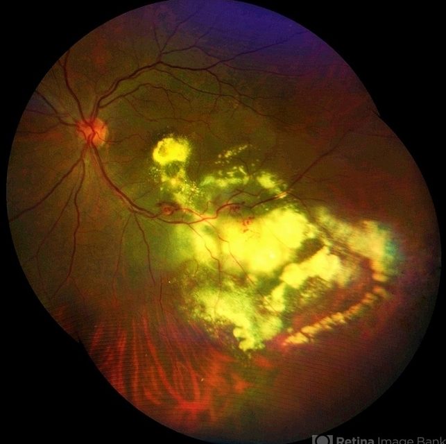
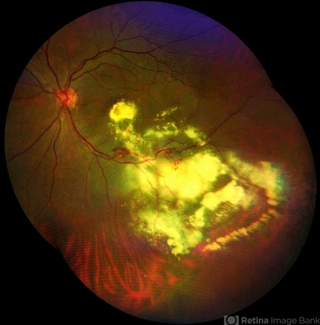
- Fundus Examination: Numerous small, round, yellowish exudates clustered around the macula and optic disc in the left eye. A few microaneurysms were noted in the mid-peripheral retina.
- Fluorescein Angiography: Multiple areas of capillary dropout and microaneurysms with late-phase leakage around the macula.
- Optical Coherence Tomography (OCT): Evidence of macular edema and intraretinal fluid in the left eye.
- Blood Pressure Measurement: 150/90 mmHg.
A diagnosis of Leber’s Miliary Aneurysm was made.
Leber’s Miliary Aneurysm(LMA) diseases entity
Leber’s Miliary Aneurysm (LMA) is a rare, congenital retinal vascular anomaly that typically presents with multiple small aneurysms and exudative retinopathy.
The condition is often unilateral and can lead to visual impairment if not managed appropriately.
This article will delve into the pathophysiology, clinical features, diagnosis, differential diagnoses, and management strategies for Leber’s Miliary Aneurysm.
Fundus Explorer Pro
Photograph the retinal findings described here with the phone already in your pocket — 22 D optics and built-in illumination in one handheld unit.
From Choroida — the team behind this sitePathophysiology
Leber’s Miliary Aneurysm (LMA) is thought to result from congenital abnormalities in the retinal vasculature. The condition involves the formation of multiple tiny aneurysms, primarily in the retinal arterioles.
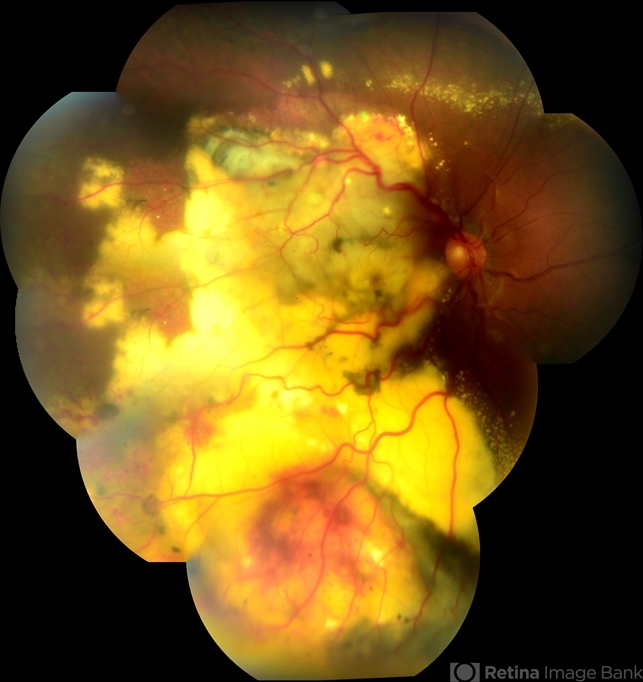
These aneurysms can lead to leakage of fluid and lipids, causing exudation and subsequent retinal thickening. Chronic leakage and exudation can lead to macular edema, lipid deposition, and fibrosis, which contribute to visual impairment.
Leber’s Miliary Aneurysm(LMA) Epidemiology
LMA is a rare condition with no clear predilection for gender or race. It typically manifests in the second to fourth decades of life. While it is generally unilateral, bilateral cases have been reported.
The prevalence of Leber’s Miliary Aneurysm (LMA) is not well-documented due to its rarity and often subtle clinical presentation.
Clinical Features
Patients with LMA may present with various symptoms, depending on the severity and location of the aneurysms:
- Blurred Vision: Commonly reported, especially if macular involvement is significant.
- Decreased Visual Acuity: Results from macular edema or lipid deposition in the central retina.
- Metamorphopsia: Distorted vision may occur if the macula is affected.
- Floaters: Less commonly, patients may report floaters due to vitreous involvement.
Examination Findings
- Fundus Examination: Characteristic multiple small, round, yellowish exudates around the macula and optic disc. Microaneurysms may be scattered throughout the retina.
- Fluorescein Angiography: Reveals areas of capillary dropout, microaneurysms, and late-phase leakage.
- Optical Coherence Tomography (OCT): Shows intraretinal fluid, macular edema, and retinal thickening.
- Systemic Evaluation: Hypertension is a common finding in patients with LMA, necessitating blood pressure monitoring.
Differential Diagnosis
The differential diagnosis for Leber’s Miliary Aneurysm (LMA) includes several other conditions with similar clinical features:
- Diabetic Retinopathy: Characterized by microaneurysms, retinal hemorrhages, and neovascularization, usually in patients with diabetes.
- Retinal Vasculitis: Inflammatory condition of the retinal vessels, often associated with systemic autoimmune diseases.
- Coats’ Disease: Unilateral retinal telangiectasia and exudation, typically presenting in children and young adults.
- Retinal Vein Occlusion: Presents with retinal hemorrhages, edema, and dilated, tortuous veins.
Leber’s Miliary Aneurysm(LMA) Diagnosis
Diagnosing Leber’s Miliary Aneurysm (LMA) involves a combination of clinical evaluation and imaging studies:
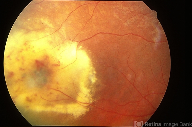
- Clinical Examination: Thorough fundus examination to identify characteristic exudates and aneurysms.
- Fluorescein Angiography: Essential for visualizing capillary dropout and leakage.
- Optical Coherence Tomography (OCT): Provides detailed imaging of retinal structures, highlighting macular edema and intraretinal fluid.
- Systemic Workup: To assess for associated conditions such as hypertension and hyperlipidemia.
Management of Leber’s Miliary Aneurysm(LMA)
Management of Leber’s Miliary Aneurysm (LMA) focuses on controlling the underlying systemic conditions and addressing the retinal pathology:
- Observation: In mild cases without significant macular involvement, regular monitoring may be sufficient.
- Laser Photocoagulation: Targeted laser treatment of the aneurysms can reduce leakage and exudation.
- Anti-VEGF Therapy: Intravitreal injections of anti-VEGF agents can help reduce macular edema and improve visual acuity.
- Systemic Management: Controlling hypertension and hyperlipidemia is crucial to prevent progression of the condition.
- Follow-Up: Regular follow-up to monitor for recurrence or progression of retinal changes.
Prognosis
The prognosis for patients with Leber’s Miliary Aneurysm (LMA) varies depending on the severity of the macular involvement and the effectiveness of treatment.
With appropriate management, many patients can achieve stabilization of their vision. However, chronic macular edema and lipid deposition can lead to permanent visual impairment in some cases.
Prevention
There are no specific preventive measures for Leber’s Miliary Aneurysm (LMA) , given its congenital nature. Public health efforts should focus on raising awareness among healthcare providers to ensure timely diagnosis and management.
Early intervention can help prevent significant visual impairment and improve outcomes for patients with Leber’s Miliary Aneurysm (LMA).

Conclusion
Leber’s Miliary Aneurysm (LMA) is a rare but significant congenital retinal vascular anomaly. Understanding its pathophysiology, clinical features, and management options is crucial for improving patient outcomes.
Advances in diagnostic imaging and therapeutic strategies offer hope for better management and prognosis for patients with Leber’s Miliary Aneurysm (LMA).
Regular monitoring and appropriate systemic management are essential components of care for individuals affected by this condition.


Document what you see
Two smartphone imaging tools built for everyday clinic use — one for the slit lamp, one for the fundus.
From Choroida — the team behind this siteReferences
- Gass, J. D. (1968). Leber’s Miliary Aneurysm. Archives of Ophthalmology, 80(2), 177-185.
- Shields, J. A., & Shields, C. L. (2001). Retina: Surgical and Medical Management. Lippincott Williams & Wilkins.
- Yannuzzi, L. A., & Freund, K. B. (2009). Retinal Vascular Disease. In Vitreoretinal Disease: The Essentials (pp. 205-215). Springer.
- Gass, J. D. M. (1996). Stereoscopic Atlas of Macular Diseases: Diagnosis and Treatment. Mosby.
- Ryan, S. J. (2006). Retina (4th ed.). Elsevier Health Sciences.
