
CASE REPORT
A 25-year-old male patient presented to an ophthalmology clinic with a chief complaint of blurred vision in both eyes. He reported that the vision had been gradually worsening over the past few months.
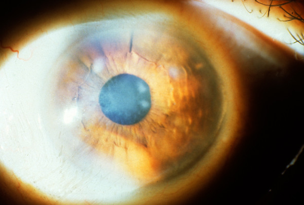
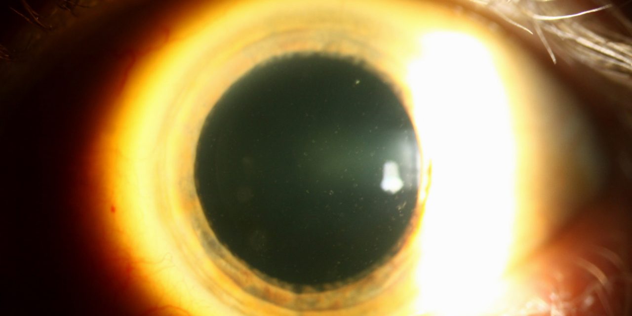
On examination, the patient was found to have multiple, small, white, opaque flecks in the corneal stroma of both eyes. The flecks were most prominent in the central and peripheral cornea.
The patient was diagnosed with Fleck Corneal Dystrophy.
DISEASE
Fleck corneal dystrophy (FCD), also known as Francois-Neetens speckled corneal dystrophy is a rare, non-inflammatory, and asymptomatic form of stromal corneal dystrophy. It is an autosomal dominant disease caused by mutations in the PIKFYVE gene on chromosome 2 (2q35).
This gene encodes for an enzyme PIKfyve also known as phosphatidylinositol-3-phosphate 5-kinase type III (PIP5k3). The PIKfyve enzyme regulates cytoskeleton structure, and membrane trafficking and has a role in the biosynthesis of endosome carrier vesicles from early endosomes in the keratocytes.
Fleck corneal dystrophy is mainly diagnosed with slit lamp by retro illumination technique. Further, image modalities such as confocal microscopy and anterior segment OCT can aid in diagnosis. In the end, the diagnosis is confirmed by histological examination.
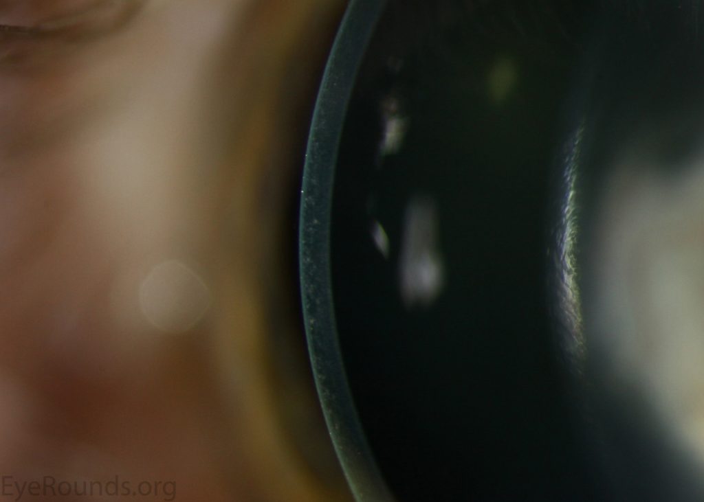
Patients are usually asymptomatic and have normal vision. The flecks of fleck dystrophy can appear as early as 2 years of age or sometimes even at birth and remains non-progressive throughout life.
All-fit Slit-Lamp Adapter
Record and share exactly what you see at the slit lamp. One adapter fits any slit lamp or surgical microscope — and any smartphone.
From Choroida — the team behind this siteClinical examination
On clinical examination, Fleck corneal dystrophy is presented as numerous tiny opacities spreading throughout the cornea. There is no change in corneal thickness in FCD.
Upon examination, There may be two types of opacities. One type consists of a small oval, round, semicircular, or wreath-like-shaped opacities with distinct borders (flecks). Another type of opacities is small greyish colored, resembling snowflakes with ill-defined margins.
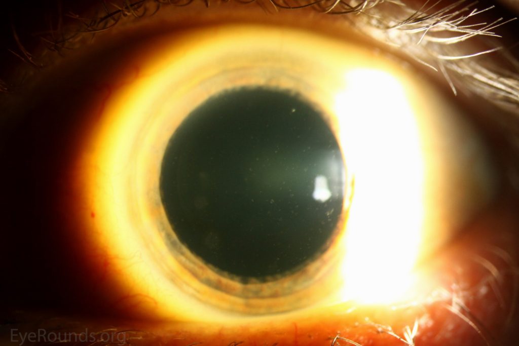
Both of these opacities are different in appearance but found symmetrically in both corneas and are observed in the same family member and even in the same individual. The stroma present between the opacities remains normal. The corneal epithelium, bowman layer, Descemet membrane, and endothelium remain normal.
MANAGEMENT
Fleck corneal dystrophy is non-progressive and asymptomatic and therefore has little clinical significance. Therefore, it usually does not require any treatment.


Document what you see
Two smartphone imaging tools built for everyday clinic use — one for the slit lamp, one for the fundus.
From Choroida — the team behind this siteREFERENCES
- Li, S., Tiab, L., Jiao, X., Munier, F. L., Zografos, L., Frueh, B. E., Sergeev, Y., Smith, J., Rubin, B., Meallet, M. A., Forster, R. K., Hejtmancik, J. F., Schorderet, D. F. Mutations in PIP5K3 are associated with Francois-Neetens mouchetee fleck corneal dystrophy. Am. J. Hum. Genet. 77: 54-63, 2005. PMID 15902656.
- Jiao, X., Munier, F. L., Schorderet, D. F., Zografos, L., Smith, J., Rubin, B., Hejtmancik, J. F. Genetic linkage of Francois-Neetens fleck (mouchetee) corneal dystrophy to chromosome 2q35. Hum. Genet. 112: 593-599, 2003. PMID 12607114
- Klintworth GK (2009). “Corneal dystrophies”. Orphanet J Rare Dis. 4: 7. doi:10.1186/1750-1172-4-7. PMC 2695576. PMID 19236704.
- Goldberg, M. F., Krimmer, B., Sugar, J., Sewell, J., Wong, P. Variable expression in flecked (speckled) dystrophy of the cornea. Ann. Ophthal. 9: 889-896, 1977. PMID: 302662
- Kawasaki, S., Yamasaki, K., Nakagawa, H., Shinomiya, K., Nakatsukasa, M., Nakai, Y., Kinoshita, S. A novel mutation (p.Glu2389AspfsX16) of the phosphoinositide kinase, FYVE finger containing gene found in a Japanese patient with fleck corneal dystrophy. Molec. Vis. 18: 2954-2960, 2012. PMID: 23288988
- Patten JT, Hyndiuk RA, Donaldson DD, Herman SJ, Ostler HB. Fleck (Mouchetée) dystrophy of the cornea. Ann Ophthalmol. 1976 Jan;8(1):25-32. PMID: 1082286.
Test yourself
A few questions straight from this article.
-
Fleck corneal dystrophy belongs to which group of corneal dystrophies?
Fleck corneal dystrophy, also called Francois-Neetens speckled corneal dystrophy, is a rare, non-inflammatory, usually asymptomatic stromal corneal dystrophy. -
Mutations in which gene cause fleck corneal dystrophy?
Fleck corneal dystrophy is caused by mutations in the PIKFYVE gene on chromosome 2 (2q35), which encodes the enzyme PIKfyve (PIP5K3). -
In fleck corneal dystrophy, the PIKfyve enzyme regulates membrane trafficking in which corneal cells?
PIKfyve regulates cytoskeleton structure and membrane trafficking and helps form endosome carrier vesicles from early endosomes in the keratocytes. -
Which slit-lamp technique is mainly used to diagnose fleck corneal dystrophy?
Fleck corneal dystrophy is mainly diagnosed at the slit lamp using the retroillumination technique. -
How is the diagnosis of fleck corneal dystrophy ultimately confirmed?
Confocal microscopy and anterior segment OCT can aid the diagnosis, but it is finally confirmed by histological examination. -
What happens to corneal thickness in fleck corneal dystrophy?
Fleck corneal dystrophy presents as numerous tiny opacities spread through the cornea, with no change in corneal thickness. -
Besides discrete, sharply bordered flecks, what other opacity occurs in fleck corneal dystrophy?
One opacity type is small oval, round, semicircular or wreath-shaped flecks with distinct borders; the other is small greyish snowflake-like opacities with ill-defined margins. -
What does the stroma between the opacities look like in fleck corneal dystrophy?
The stroma between the opacities remains normal, as do the epithelium, Bowman layer, Descemet membrane and endothelium. -
How are the two opacity types of fleck corneal dystrophy distributed?
Though different in appearance, both opacity types are found symmetrically in both corneas, within the same family and even the same individual. -
What treatment does fleck corneal dystrophy usually require?
Because fleck corneal dystrophy is non-progressive and asymptomatic, it has little clinical significance and usually needs no treatment.
