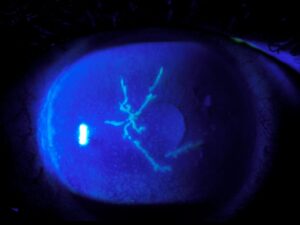
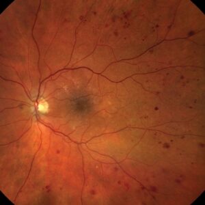
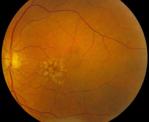
Acanthamoeba stages describe how this contact lens keratitis moves from the epithelium into the stroma and beyond.
The organism is a free-living protozoan found in tap water, soil and lens cases. Because early signs look mild, many patients first receive a herpes or bacterial diagnosis, so recognizing the stage at presentation helps you predict outcome and plan therapy.
Why Acanthamoeba Stages Matter
Depth of invasion largely determines how well the eye responds to treatment. Epithelial disease often clears with topical biguanides and leaves little scarring. In contrast, stromal disease needs longer therapy and carries a higher risk of surgery, and several series also link diagnostic delay with worse visual outcomes.
Symptoms Across Acanthamoeba Stages
Pain is often severe and seems out of proportion to the signs, and patients also describe photophobia, tearing and blurred vision. However, pain varies, and some early cases report only mild discomfort. As a result, the absence of severe pain should never exclude the diagnosis.
All-fit Slit-Lamp Adapter
Record and share exactly what you see at the slit lamp. One adapter fits any slit lamp or surgical microscope — and any smartphone.
From Choroida — the team behind this siteStage 1: Epitheliopathy
The earliest stage involves the corneal epithelium alone. At the slit lamp, you may see punctate keratopathy, microcystic edema or irregular epithelial haze. Raised epithelial lines and pseudodendrites can also appear, and these pseudodendrites lack the terminal bulbs of herpetic dendrites and stain less crisply with fluorescein.
Pseudodendrites in Early Acanthamoeba Stages
Misreading pseudodendrites as herpes simplex keratitis is a classic error, and antivirals, and sometimes steroids, then delay the correct treatment. A contact lens wearer with a “dendrite” and no herpes history also deserves suspicion. Corneal scraping for culture, PCR and confocal microscopy can confirm the diagnosis at this stage.
Stage 2: Radial Keratoneuritis
Next, trophozoites track along corneal nerves and produce perineural infiltrates, which appear as fine linear or beaded lines radiating from the center toward the limbus. The sign is fairly specific for Acanthamoeba and often coincides with intense pain (see radial keratoneuritis), and because it appears early, it offers a valuable diagnostic window.
Stromal Invasion in Acanthamoeba Stages
Stromal involvement begins with patchy anterior infiltrates under an intact or irregular epithelium, and epithelial breakdown may follow as stromal edema increases. At this point, cysts lodge deeper in the stroma, where drugs penetrate less well. Consequently, treatment becomes longer and relapse becomes more likely.
Stage 3: Ring Infiltrate
The classic ring infiltrate forms a partial or complete annulus in the paracentral stroma, which reflects an immune response to amoebic antigens and signals established stromal disease. Many textbook photos show this ring, while many eyes never develop it. Therefore, waiting for a ring before testing guarantees a late diagnosis (see ring infiltrate).
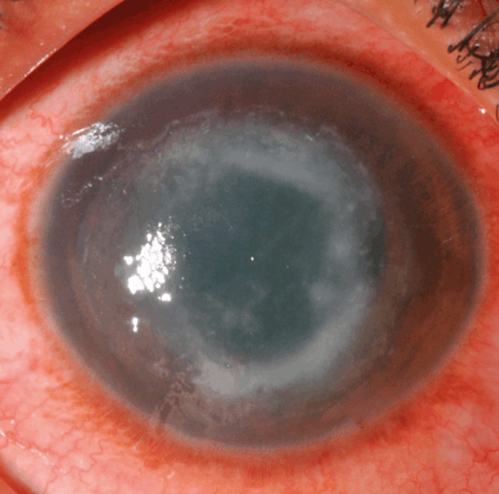
Advanced Acanthamoeba Stages
Without effective therapy, the stroma can ulcerate and thin progressively, and secondary bacterial or fungal infection may also complicate the ulcer. Descemetocele and perforation then become real risks, and at this stage patients may need therapeutic keratoplasty, although grafts carry a recurrence risk. Persistent inflammation may also cause cataract, secondary glaucoma, iris atrophy and a fixed dilated pupil.
Limbitis and Scleritis
Some patients develop limbitis and nodular or diffuse anterior scleritis, which clinicians usually regard as immune mediated because organisms rarely appear in scleral tissue. Scleritis causes severe pain and often needs systemic anti-inflammatory therapy alongside antiamoebic drugs, and it marks severe disease with a guarded prognosis.
Acanthamoeba Stages and Prognosis
Early epithelial disease generally carries the best visual prognosis, while ring infiltrate, deep stromal disease and scleritis predict longer treatment and more surgery. For example, a large UK series associated advanced disease at presentation with poorer outcomes, and several reports also associate steroid use before antiamoebic therapy with worse results.
Treatment by Acanthamoeba Stages
Topical biguanides form the backbone of therapy at every stage, with options that include polihexanide or chlorhexidine, often combined with a diamidine such as propamidine. The ODAK trial supported high-concentration polihexanide monotherapy as an alternative to dual therapy (see polihexanide), and treatment usually lasts months, with deeper stages needing longer courses.
Steroids in Later Acanthamoeba Stages
Steroids do not kill cysts, and they may worsen infection if started too early. However, some specialists add them cautiously for scleritis or severe inflammation once antiamoebic therapy is established, and then the patient needs close review because relapse can occur.
Counseling the Patient
Tell the patient that the infection is treatable, but recovery is slow, and stress strict avoidance of water contact with lenses during and after treatment. Patients with stromal disease should understand that surgery or scarring remains possible, and you should discuss lens hygiene before any return to wear.
Pitfalls to Avoid
- Do not label every contact lens pseudodendrite as herpes
- Never wait for a ring infiltrate before scraping
- Avoid steroids before starting antiamoebic therapy
- Do not stop drops early once symptoms improve
- Check pressure and lens status in chronic cases


Document what you see
Two smartphone imaging tools built for everyday clinic use — one for the slit lamp, one for the fundus.
From Choroida — the team behind this siteReferences
- Dart JKG, Saw VPJ, Kilvington S. Acanthamoeba keratitis: diagnosis and treatment update 2009. Am J Ophthalmol. 2009;148:487-499.
- Claerhout I, Goegebuer A, Van Den Broecke C, Kestelyn P. Delay in diagnosis and outcome of Acanthamoeba keratitis. Graefes Arch Clin Exp Ophthalmol. 2004;242:648-653.
- Carnt N, Robaei D, Minassian DC, Dart JKG. Acanthamoeba keratitis in 194 patients: risk factors for bad outcomes and severe inflammatory complications. Br J Ophthalmol. 2018;102:1431-1435.
- Dart JKG, Papa V, Rama P, et al. The Orphan Drug for Acanthamoeba Keratitis (ODAK) trial: PHMB 0.08% (polihexanide) and placebo versus PHMB 0.02% and propamidine 0.1%. Ophthalmology. 2024;131:277-287.
Test yourself
A few questions straight from this article.
-
In Acanthamoeba keratitis, which corneal layer is involved alone in stage 1 disease?
The article states the earliest stage, epitheliopathy, involves the corneal epithelium alone, with signs such as punctate keratopathy, microcystic edema or epithelial haze. -
How do pseudodendrites of Acanthamoeba keratitis differ from herpetic dendrites on slit lamp examination?
The article notes Acanthamoeba pseudodendrites lack the terminal bulbs of herpetic dendrites and stain less crisply with fluorescein, which helps avoid misdiagnosis as herpes. -
In Acanthamoeba keratitis, what typically produces the radial keratoneuritis seen in stage 2?
Trophozoites track along corneal nerves and produce perineural infiltrates, seen as fine linear or beaded lines radiating toward the limbus. The sign is fairly specific for Acanthamoeba. -
What does the classic ring infiltrate in Acanthamoeba keratitis reflect?
The article says the partial or complete paracentral ring reflects an immune response to amoebic antigens and signals established stromal disease. -
Why should clinicians not wait for a ring infiltrate before testing for Acanthamoeba keratitis?
Textbook photos show the ring, but many eyes never develop it. Waiting for one before testing guarantees a late diagnosis. -
A contact lens wearer has a dendrite but no herpes history. Which condition does the article say deserves suspicion?
The article states a contact lens wearer with a dendrite and no herpes history deserves suspicion for Acanthamoeba, since misreading pseudodendrites as herpes is a classic error. -
Which topical agents form the backbone of Acanthamoeba keratitis therapy at every stage?
Topical biguanides, including polihexanide or chlorhexidine, are the backbone of therapy at every stage, often combined with a diamidine such as propamidine. -
What did the ODAK trial support in Acanthamoeba keratitis treatment?
The article states the ODAK trial supported high-concentration polihexanide monotherapy as an alternative to dual therapy. -
Why can starting steroids too early be harmful in Acanthamoeba keratitis?
Steroids do not kill cysts and may worsen infection if started too early. Some specialists add them cautiously for scleritis or severe inflammation once antiamoebic therapy is established. -
In Acanthamoeba keratitis, why is scleritis usually regarded as immune mediated?
Clinicians usually regard limbitis and scleritis as immune mediated because organisms rarely appear in scleral tissue. Scleritis marks severe disease with a guarded prognosis and often needs systemic anti-inflammatory therapy.

Writes and reviews the clinical articles on Educate. Every post is checked against peer-reviewed sources before it goes live.