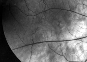
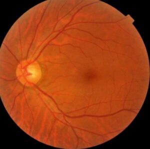
Preeclampsia retinopathy describes the retinal changes that occur in pregnant patients with severe hypertension and preeclampsia.
Many patients have visual symptoms, and some have serious retinal findings that signal severe disease.
Ophthalmologists should know the signs, since they may help the obstetric team to judge the stage of illness.
What Preeclampsia Retinopathy Is
Preeclampsia is a hypertensive disorder that appears after 20 weeks of pregnancy, with proteinuria or organ dysfunction.
The retinal circulation reacts to the sudden rise in pressure with vasospasm, ischemia, and breakdown of the barrier.
These changes resemble those of malignant hypertension, but they develop faster and often reverse after delivery (see hypertensive retinopathy).
Symptoms of Preeclampsia Retinopathy
Patients report blurred vision, scotomas, flashes, photophobia, or transient loss of vision.
Some develop sudden severe loss from retinal detachment, retinal artery occlusion, or cortical changes.
Roos and colleagues reviewed visual disturbances in preeclampsia and eclampsia and found them in a substantial share of severe cases.
Fundus Explorer Pro
Photograph the retinal findings described here with the phone already in your pocket — 22 D optics and built-in illumination in one handheld unit.
From Choroida — the team behind this siteSigns of Preeclampsia Retinopathy
- Generalized or focal arteriolar narrowing
- Flame hemorrhages and cotton wool spots
- Retinal edema and hard exudates
- Serous retinal detachment in severe disease
Optic disc swelling can occur and suggests a hypertensive emergency (see exudative retinal detachment).
Choroidal Involvement
The choroid also suffers, and this explains the serous detachment.
Focal areas of RPE necrosis, known as Elschnig spots, may appear after the detachment resolves.
Fluorescein angiography shows patchy choroidal filling and leakage at the level of the RPE.
OCT shows subretinal fluid with an intact retina above it.
Management of Preeclampsia Retinopathy
Control of blood pressure and delivery treat the cause, and the obstetric team leads this process.
Ophthalmologists document findings and monitor the retina until it settles.
Do not treat the detachment with laser or injections, since it usually resolves after delivery.
Counsel patients that most recover vision within weeks, although some keep scars or defects (see diabetic retinopathy in pregnancy).
Differential Diagnosis of Preeclampsia Retinopathy
Consider central serous chorioretinopathy, which also affects pregnant patients.
Also consider Purtscher-like retinopathy, HELLP syndrome, and thrombotic microangiopathy.
The history, the blood pressure, and the laboratory tests help to separate them (see Purtscher retinopathy).
An Example Case
A 29-year-old woman at 34 weeks of pregnancy has a blood pressure of 170 over 110 and sudden blurred vision.
Examination shows narrowed arterioles, a few flame hemorrhages, and a serous detachment in the left eye.
The obstetric team controls her blood pressure and delivers the baby the same day.
Within three weeks, the fluid resolves, and she regains 20/25 vision, with faint pigment changes in the left macula.
Questions Patients Ask
Patients ask whether the vision loss is permanent, and most recover after delivery.
They also ask whether the eye disease will return in another pregnancy, and the risk depends on the recurrence of preeclampsia.
Because mothers worry, explain the good outlook and the follow-up plan.
Pitfalls to Avoid
- Treating serous detachment with laser during pregnancy
- Delaying referral to the obstetric team when findings suggest severe disease
- Missing optic disc swelling that signals a hypertensive emergency
- Forgetting to check for Purtscher-like changes in HELLP syndrome
Looking Ahead
Retinal imaging may help to predict preeclampsia, but studies are small.
Also, OCT may track choroidal changes during treatment.
However, the main role of the eye exam stays supportive.
Practical Points
Dilate the pupil safely, and document findings with photographs.
Then communicate results to the obstetrician the same day.
Also arrange a follow-up exam within weeks after delivery.
Imaging in Preeclampsia Retinopathy
Fundus photographs document hemorrhages, exudates, and detachment.
OCT shows subretinal fluid, and it measures the response after delivery.
Fluorescein angiography helps in unclear cases, though many centers avoid it in pregnancy unless needed.
Therefore, rely on OCT for follow-up.
Long-Term Effects
Most eyes recover fully.
However, some patients keep areas of RPE atrophy or Elschnig spots.
Also, patients with preeclampsia face higher risks of later hypertension and cardiovascular disease, so advise regular medical review.
Finally, remind patients to report new visual symptoms in later pregnancies.
Collaboration With the Obstetric Team
Send a short note that gives the findings, the severity, and the follow-up plan.
Because the team makes urgent decisions, communicate the same day.
Then confirm the plan after delivery.
Counselling Before Discharge
Explain the findings in simple language and give written warning signs.
Then arrange a visit within two to four weeks after delivery.
Because many mothers feel overwhelmed, involve a family member in the discussion.
Also give a number to call for new symptoms.


Document what you see
Two smartphone imaging tools built for everyday clinic use — one for the slit lamp, one for the fundus.
From Choroida — the team behind this siteReferences
- Roos NM, Wiegman MJ, Jansonius NM, Zeeman GG. Visual disturbances in (pre)eclampsia. Obstet Gynecol Surv. 2012;67:242-250.
- Schultz KL, Birnbaum AD, Goldstein DA. Ocular disease in pregnancy. Curr Opin Ophthalmol. 2005;16:308-314.
- Abu Samra K. The eye and visual system in the preeclampsia/eclampsia syndrome: what to expect? Saudi J Ophthalmol. 2013;27:51-53.
- American Academy of Ophthalmology. Basic and Clinical Science Course, Section 12: Retina and Vitreous. AAO; current edition.

Writes and reviews the clinical articles on Educate. Every post is checked against peer-reviewed sources before it goes live.