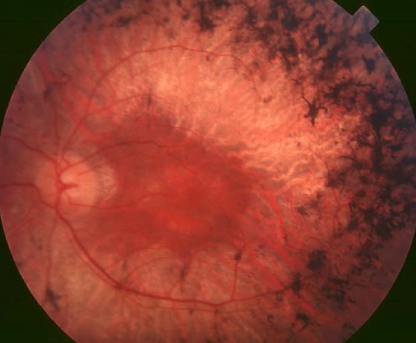
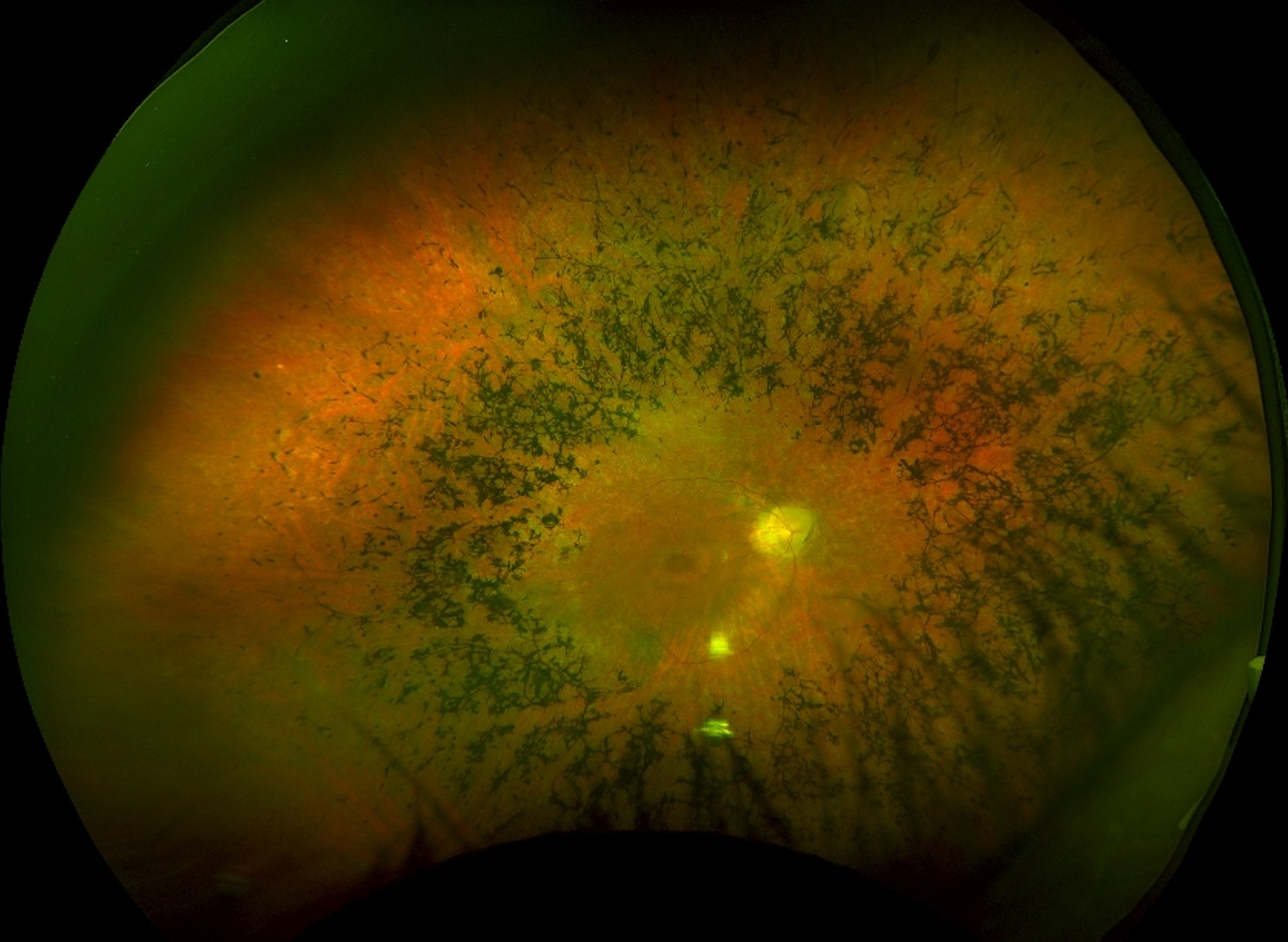
Bone spicule pigmentation describes dark, branching clumps of pigment that sit in the inner retina along the vessels.
It is the classic fundus sign of retinitis pigmentosa, but several other diseases leave the same mark.
Therefore, the sign alone never confirms the diagnosis.
What Bone Spicule Pigmentation Looks Like
Clumps of pigment form in the midperiphery first, then spread toward the posterior pole and the far periphery.
They are black and angular, and they often gather around retinal veins.
Also, the retinal arterioles look narrow, and the optic disc may show waxy pallor (see retinitis pigmentosa).
Between the clumps, the retinal pigment epithelium looks atrophic, and the choroidal vessels may show through.
How Bone Spicule Pigmentation Develops
When photoreceptors die, the retinal pigment epithelium loses contact with the outer retina.
Then the pigment cells migrate into the inner retina and wrap around the vessels.
Hartong and colleagues described this as a late event that follows rod loss.
As a result, pigment appears only after substantial photoreceptor damage has occurred.
Fundus Explorer Pro
Photograph the retinal findings described here with the phone already in your pocket — 22 D optics and built-in illumination in one handheld unit.
From Choroida — the team behind this siteGenetic Causes of Bone Spicule Pigmentation
- Nonsyndromic retinitis pigmentosa, which is the most common cause
- Usher syndrome, with congenital hearing loss (see Usher syndrome)
- Bardet-Biedl syndrome, with obesity, polydactyly, and renal disease
- Late Leber congenital amaurosis and some cone-rod dystrophies
Check the family tree, since dominant, recessive, and X-linked patterns all occur.
Acquired Causes of Bone Spicule Pigmentation
Old inflammation can mimic the picture.
Congenital rubella and syphilis both leave pigmentary change, and a treated retinal detachment can leave similar pigment (see ocular syphilis).
Also consider toxic causes such as thioridazine and chloroquine, and a retained iron foreign body.
Unilateral or very asymmetric disease points to an acquired cause, for example diffuse unilateral subacute neuroretinitis.
Examination and Tests for Bone Spicule Pigmentation
Ask about night vision, peripheral field loss, hearing, and family history.
Also record drug exposure, past infection, and any eye injury.
The electroretinogram shows reduced rod and cone responses in retinitis pigmentosa, and it may become nonrecordable in advanced disease (see electroretinography).
Visual fields show a ring scotoma that narrows over time.
Optical coherence tomography often shows loss of the ellipsoid zone outside a preserved central island.
Fundus autofluorescence shows a ring of increased signal around the macula.
Management and Counselling for Bone Spicule Pigmentation
Confirm the genetic diagnosis with a gene panel, because it guides counseling and trial access (see gene therapy).
Treat cystoid macular edema with carbonic anhydrase inhibitors when it occurs.
Also remove visually significant posterior subcapsular cataract.
Refer to low vision services early, and discuss driving and mobility.
Because the family may be at risk, offer genetic counseling to relatives.
When Bone Spicule Pigmentation Is Not Retinitis Pigmentosa
Some patients have pigment clumps but no family history and no night blindness.
Often the findings are unilateral, or the two eyes differ greatly.
This picture suggests pseudoretinitis pigmentosa, and the cause is usually acquired (see unilateral pigmentary retinopathy).
Causes include old inflammation, trauma, resolved detachment, retained iron, and diffuse unilateral subacute neuroretinitis.
Therefore, obtain old records, order serology, and compare the electroretinogram in both eyes.
Order orbital imaging when you suspect a foreign body.
Treatable Causes of Bone Spicule Pigmentation
A few systemic diseases produce the same retinal picture and respond to treatment.
Refsum disease causes pigmentary retinopathy with anosmia, ataxia, and neuropathy, and a low phytanic acid diet helps.
Abetalipoproteinemia causes it through vitamin A and E deficiency, and supplements slow the damage.
Also check vitamin A levels in patients with malabsorption.
Because these conditions are rare, a simple blood test and a short history are enough to exclude them.
Practical Points at the Bedside
Compare the two eyes first, because symmetry points to a hereditary disease.
Then check the disc, the arterioles, and the macula for edema or an epiretinal membrane.
Also look at the lens, since posterior subcapsular cataract is common in retinitis pigmentosa.
Take photographs, so that change can be judged on later visits.
Referral and Follow-Up
Refer to a retinal dystrophy service when the pattern is bilateral and progressive.
Also arrange hearing tests when Usher syndrome is possible.
Review the patient yearly with fields and optical coherence tomography.
Because progress varies, give the patient a clear contact for new symptoms.
Imaging Clues That Help
Optical coherence tomography can separate retinitis pigmentosa from acquired mimics.
Look for a thinned outer nuclear layer and a preserved central ellipsoid zone in dystrophy.
Also check for cystoid spaces, because they respond to treatment.
In acquired disease, the damage often follows the area of old inflammation or trauma.
For the latest studies, see the PubMed results for bone spicule pigmentation.


Document what you see
Two smartphone imaging tools built for everyday clinic use — one for the slit lamp, one for the fundus.
From Choroida — the team behind this siteReferences
- Hartong DT, Berson EL, Dryja TP. Retinitis pigmentosa. Lancet. 2006;368:1795-1809.
- Verbakel SK, van Huet RAC, Boon CJF, et al. Non-syndromic retinitis pigmentosa. Prog Retin Eye Res. 2018;66:157-186.
- Hamel C. Retinitis pigmentosa. Orphanet J Rare Dis. 2006;1:40.
- Berson EL. Retinitis pigmentosa. The Friedenwald Lecture. Invest Ophthalmol Vis Sci. 1993;34:1659-1676.

Writes and reviews the clinical articles on Educate. Every post is checked against peer-reviewed sources before it goes live.