Tuberous sclerosis complex is an autosomal dominant disorder caused by mutations in TSC1 or TSC2, which activate the mTOR pathway and produce benign tumors in many organs.
The eye is also involved in about half of patients.
The retinal findings can also help to make the diagnosis in a child with seizures or skin changes.
Eye findings are usually asymptomatic, and the main task for the ophthalmologist is also to recognize them and refer.
Retinal Astrocytic Hamartomas in Tuberous Sclerosis
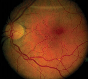
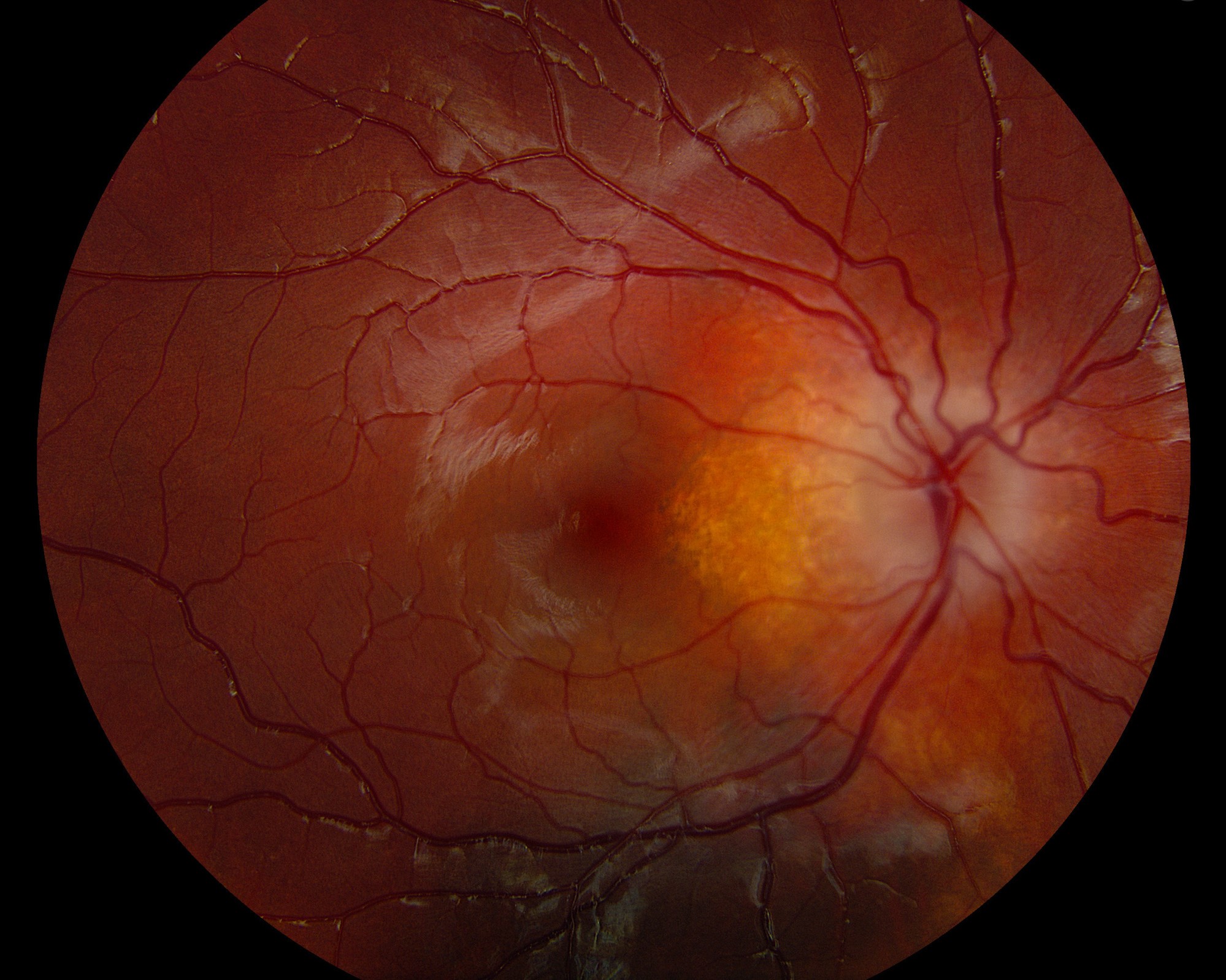
These benign tumors of glial origin arise in the nerve fiber layer and appear in about 30 to 50 percent of patients (see the references).
There are three morphologies:
- Flat, translucent lesions with ill-defined borders, the most common
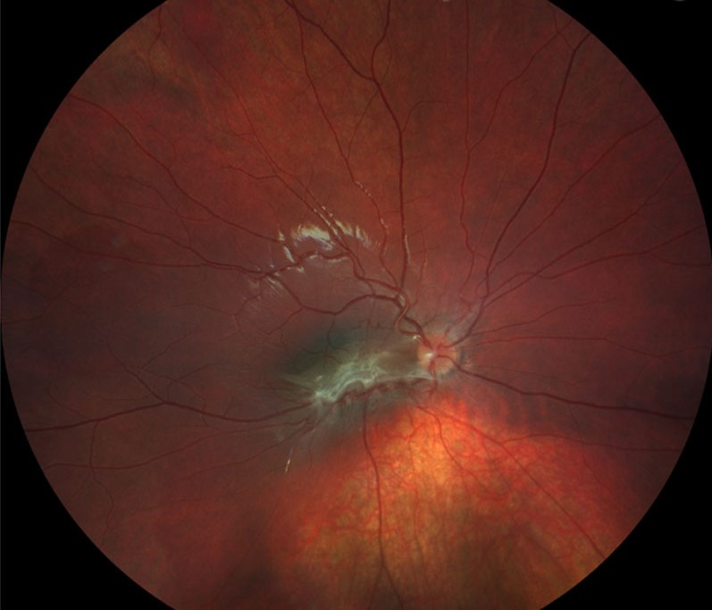
- Mulberry lesions, which are also elevated and calcified, and often near the disc
- Transitional lesions, with a mix of both
They may also be single or multiple, and unilateral or bilateral (see retinal astrocytic hamartoma).
Tuberous Sclerosis: Other Eye Findings
- Retinal achromic patches, areas of depigmentation in the periphery
- Eyelid angiofibromas, which are part of the facial rash
- Hypopigmented iris spots
- Optic disc drusen and, rarely, papilledema from a brain tumor
- Strabismus and visual field defects from brain lesions
Fundus Explorer Pro
Photograph the retinal findings described here with the phone already in your pocket — 22 D optics and built-in illumination in one handheld unit.
From Choroida — the team behind this siteComplications of Tuberous Sclerosis
Most hamartomas do not grow, but a few enlarge and cause exudation, hemorrhage, or retinal detachment, and a vitreous hemorrhage can reduce vision.
Also, rapid growth is more common in young children, and any change in appearance needs close follow-up and sometimes treatment with laser, photodynamic therapy, anti-VEGF drugs, or surgery.
Systemic Features
The skin shows hypomelanotic macules, facial angiofibromas, shagreen patches, and ungual fibromas.
Also, brain lesions include cortical tubers, subependymal nodules, and giant cell astrocytomas, which cause seizures, developmental delay, and autism.
In addition, other tumors include cardiac rhabdomyomas, renal angiomyolipomas, and lung lymphangioleiomyomatosis in women (see the references).
Diagnostic Criteria
The 2012 international criteria require two major features or one major and two minor features for a definite diagnosis.
A genetic mutation is also sufficient.
The criteria also list multiple retinal hamartomas as a major feature, and retinal achromic patches as a minor one.
Differential Diagnosis of Tuberous Sclerosis
Astrocytic hamartomas can look like retinoblastoma in a child, and calcified lesions need careful imaging (see retinoblastoma).
Other phakomatoses have different retinal tumors, such as the capillary hemangioblastomas of von Hippel-Lindau disease and the combined hamartomas of neurofibromatosis (see von Hippel-Lindau disease and neurofibromatosis type 1).
Management and Follow-Up for Tuberous Sclerosis
Children should have a dilated examination at diagnosis and then regularly, since new lesions can appear.
Systemic treatment includes everolimus, an mTOR inhibitor, for giant cell astrocytomas and renal tumors, and antiseizure drugs.
It has been shown to reduce some other manifestations.
Also, genetic counselling helps families to understand recurrence risks and to screen relatives.
An Example Case
A neurologist sends a 3-year-old girl with infantile spasms for an eye examination.
Examination shows a flat, translucent lesion near the optic disc in the right eye, with a calcified mulberry lesion in the left.
Dermatology had already noted hypomelanotic macules, and an MRI shows cortical tubers, which together meet the criteria for a definite diagnosis.
The team photographs the retinal lesions and reviews her every six months, with no growth over three years.
Pitfalls
- Treating a retinal hamartoma as a retinoblastoma, with unnecessary enucleation in a child
- Forgetting to examine a parent, since many have mild disease
- Stopping eye follow-up because the lesions look stable
- Missing papilledema from a brain tumor, which needs urgent imaging
Counselling Families
Explain that the retinal lesions rarely affect vision and usually stay stable, that vision problems are more often related to the brain, and that regular visits are the best protection.
Doctors advise genetic counselling, since the risk to each child of an affected parent is 50 percent, and many cases come from new mutations.
Eye Care Within a Team
Children with this condition are managed by neurology, nephrology, cardiology, dermatology, and genetics, and the ophthalmologist supports the diagnosis and watches for complications.
Share the photographs and a written report, and tell the team when a lesion changes.
Looking Ahead
Studies of mTOR inhibitors report shrinkage of some astrocytic hamartomas, and topical and systemic forms are being tested.
Early genetic diagnosis in infants is changing surveillance, since treatment can start before seizures appear.
When to Refer
Refer any infant with unexplained seizures and a retinal lesion for neurologic and genetic assessment, and refer any child with a growing hamartoma to a retina specialist.
Offer a systemic evaluation to adults with a lesion and a family history.


Document what you see
Two smartphone imaging tools built for everyday clinic use — one for the slit lamp, one for the fundus.
From Choroida — the team behind this siteReferences
- Rowley SA, O’Callaghan FJ, Osborne JP. Ophthalmic manifestations of tuberous sclerosis: a population based study. Br J Ophthalmol. 2001;85:420-423.
- Northrup H, Krueger DA. Tuberous sclerosis complex diagnostic criteria update: recommendations of the 2012 International Tuberous Sclerosis Complex Consensus Conference. Pediatr Neurol. 2013;49:243-254.
- Henske EP, Jozwiak S, Kingswood JC, Sampson JR, Thiele EA. Tuberous sclerosis complex. Nat Rev Dis Primers. 2016;2:16035.
- American Academy of Ophthalmology. Basic and Clinical Science Course, Section 12: Retina and Vitreous. AAO; current edition.

Writes and reviews the clinical articles on Educate. Every post is checked against peer-reviewed sources before it goes live.