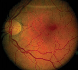
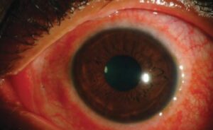
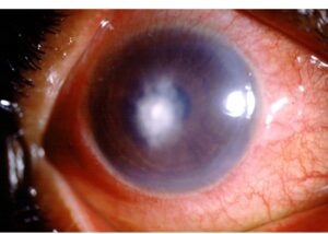
Thioridazine retinopathy is a toxic pigmentary retinopathy caused by high doses of the antipsychotic drug thioridazine.
It is rare today, because the maximum dose is limited and the drug has been largely replaced, but it remains a classic example of dose-related retinal toxicity.
Knowing the pattern helps to recognize it in patients who still take the drug.
What Is Thioridazine Retinopathy?
Thioridazine is a phenothiazine once widely used for schizophrenia.
At high doses, it accumulates in the uveal tract and the retinal pigment epithelium, which have a high content of melanin, and damages them.
Also, the risk is tied to the daily dose and to the duration of treatment, and doctors see retinopathy mostly with doses above 800 milligrams a day.
The regulators limited the maximum dose to 800 milligrams a day, and later regulators restricted the drug because of concerns about cardiac effects.
Other phenothiazines, such as chlorpromazine, cause deposits in the cornea and lens, but retinopathy is much less common with them.
Clinical Course
The effects appear in stages.
- Early stage: blurred vision, reduced night vision, and a brownish discoloration of vision, sometimes noticed within weeks of high doses
- Fundus changes: granular pigment stippling in the posterior pole and midperiphery, with later patches of nummular pigment loss or atrophy, a coin-like pattern
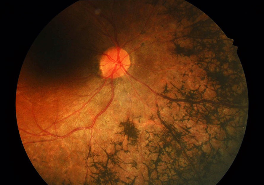
- Late stage: widespread loss of the pigment epithelium and choriocapillaris, with bone-spicule-like pigment and a resemblance to retinitis pigmentosa (see retinitis pigmentosa)
- Visual field: ring scotomas or constriction, depending on the extent
- Electroretinogram: reduced responses in advanced cases (see electroretinography)
Some patients continue to lose vision after the drug is stopped, and the damage may progress for months or longer.
Fundus Explorer Pro
Photograph the retinal findings described here with the phone already in your pocket — 22 D optics and built-in illumination in one handheld unit.
From Choroida — the team behind this sitePathology
Clinical and ultrastructural studies have shown damage to the pigment epithelium, with loss of melanin granules and degeneration of photoreceptors.
Also, the drug binds to melanin, which explains the selective effect on the pigmented tissues, and the damage seems to depend on the amount of drug that accumulates (see the references).
Risk Factors of Thioridazine Retinopathy
- Doses above the recommended maximum
- Long duration of treatment at high dose
- Other drugs that raise the level, or impaired metabolism of the drug
- Preexisting retinal disease, perhaps
Most reported cases involved daily doses above 800 milligrams, whereas cases at lower doses are rare.
Differential Diagnosis of Thioridazine Retinopathy
- Retinitis pigmentosa and other pigmentary retinopathies (see other causes of night vision loss)
- Chloroquine and hydroxychloroquine retinopathy, with a bull’s-eye pattern (see hydroxychloroquine retinopathy screening)
- Pentosan polysulfate maculopathy and other drug toxicities (see pentosan polysulfate maculopathy)
- Siderosis and other toxic retinopathies (see siderosis bulbi)
- Inherited and inflammatory retinal degenerations
A drug history clarifies the picture.
Management and Prevention for Thioridazine Retinopathy
- Stop the drug or reduce the dose, but only in consultation with the psychiatrist
- Then document the findings with photographs, fundus autofluorescence, OCT, and visual fields
- Follow the patient, since progression may occur even after cessation
- Educate prescribers and patients about the dose limit and the symptoms to report
- Consider alternative antipsychotic drugs, since they have a different safety profile
Patients on high-dose phenothiazines should also have baseline and periodic eye examinations, according to local policy.
Counselling
Explain that the retinal damage is caused by the drug, that it may continue to evolve for some time, and that early reporting of vision problems allows a change in treatment.
Also, coordinate with the mental health team, and make sure that the patient does not stop medication abruptly without advice.
Historical Context
Thioridazine was one of the most prescribed antipsychotics in the 1960s, and reports of pigmentary retinopathy appeared soon after.
In addition, doctors recognized the dose-related nature of the toxicity, and manufacturers set a ceiling on the daily dose.
Because the drug can also prolong the QT interval of the heart, many countries now reserve it for patients who have not responded to other drugs, and its use has declined.
Other Antipsychotics and the Eye
Chlorpromazine and related phenothiazines can cause deposits in the lens and cornea and skin pigmentation at high cumulative doses, and these are usually harmless to vision.
Newer atypical antipsychotics have not been linked to retinal toxicity in a clear way, though reports describe cataract and other effects with some.
In addition, clinicians should be aware of the full list of drugs a patient takes.
What to Check at Examination
- Visual acuity, color vision, and night vision
- Visual fields, looking for ring scotomas
- Fundus examination and autofluorescence, which can also show the nummular pattern of pigment loss (see fundus autofluorescence)
- OCT of the macula, for outer retinal damage
- Electroretinography, if the diagnosis is uncertain
Patients may not report symptoms because of their illness, so a systematic approach is important.
Communicating With the Psychiatrist
Send a clear letter describing the findings, the likely link to the drug, and the recommended action, and ask for a treatment review.
Avoid advising the patient to stop the drug abruptly, since relapse of the psychiatric illness is also a serious risk.


Document what you see
Two smartphone imaging tools built for everyday clinic use — one for the slit lamp, one for the fundus.
From Choroida — the team behind this siteReferences
- Miller FS III, Bunt-Milam AH, Kalina RE. Clinical-ultrastructural study of thioridazine retinopathy. Ophthalmology. 1982;89:1478-1488.
- Yanoff M, Duker JS, eds. Ophthalmology. 5th ed. Elsevier; 2019.
- Ryan SJ, Schachat AP, Wilkinson CP, et al, eds. Retina. 6th ed. Elsevier; 2018.
- American Academy of Ophthalmology. Basic and Clinical Science Course, Section 12: Retina and Vitreous. AAO; current edition.

Writes and reviews the clinical articles on Educate. Every post is checked against peer-reviewed sources before it goes live.