Open-angle vs angle-closure glaucoma is the first distinction to make in any patient with raised pressure.
The two forms differ in mechanism, risk groups and treatment, and gonioscopy separates them reliably.
Mechanism of Open-Angle Glaucoma
The drainage angle is open, but outflow is reduced because resistance sits in the trabecular meshwork. Pressure rises slowly over years, therefore it is usually silent (see primary open-angle glaucoma).
Mechanism of Angle-Closure Glaucoma
The iris blocks the trabecular meshwork, and pupillary block is the commonest cause. Plateau iris and lens factors also contribute, so pressure may rise suddenly.
All-fit Slit-Lamp Adapter
Record and share exactly what you see at the slit lamp. One adapter fits any slit lamp or surgical microscope — and any smartphone.
From Choroida — the team behind this siteOpen-Angle vs Angle-Closure: Who Is at Risk
Open-angle disease is common in older adults and people of African ancestry, while angle closure is more common in Asian populations and hyperopic eyes. Women are affected more often, and family history also matters in both.
Open-Angle vs Angle-Closure: Symptoms
Open-angle glaucoma causes no symptoms until late, while acute angle closure causes severe pain, halos and vomiting. Subacute episodes cause brief blur and headaches, so ask about halos around lights.
Open-Angle vs Angle-Closure: Signs
Open-angle eyes have normal-looking anterior chambers and open angles, while angle-closure eyes have shallow chambers and narrow angles. Acute attacks show a fixed mid-dilated pupil and corneal edema, so check the chamber depth in every patient.
Open-Angle vs Angle-Closure: Why Gonioscopy Decides
Gonioscopy views the angle structures directly and is the reference test for separating the two forms. Without it, closure may be missed, so repeat it as the lens changes with age.
Imaging Open-Angle vs Angle-Closure Eyes
Anterior segment OCT shows angle width without contact, while ultrasound biomicroscopy shows the ciliary body. These tools help but do not replace gonioscopy, so combine them with clinical examination.
Open-Angle vs Angle-Closure: Optic Nerve
Both forms cause cupping and rim loss, although in acute closure the disc may be swollen at first. Chronic closure causes slow cupping, so disc findings do not separate them.
Treatment of Open-Angle Glaucoma
Start with drops such as prostaglandin analogues, and add laser trabeculoplasty or surgery as needed. The goal is to lower pressure to target, so review regularly (see glaucoma eye drops).
Treatment of Angle-Closure Glaucoma
Laser peripheral iridotomy relieves pupillary block, and lens extraction is increasingly used. Acute attacks need medical therapy first, therefore treat the cause as well as the pressure.
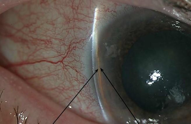
Angle Closure Suspects
Narrow angles without damage are called suspects, and the ZAP trial suggested low progression risk. Many clinicians still treat selected eyes (see primary angle closure suspect), so discuss options with the patient.
Dilation in Open-Angle vs Angle-Closure Eyes
Dilation can trigger an attack in narrow angles, so assess the angle before dilating. Warn patients about the symptoms of acute closure, and also give them an emergency contact.
Pitfalls to Avoid
- Do not dilate before checking the angle
- Never treat all raised pressure as open-angle disease
- Avoid skipping gonioscopy
- Do not forget to repeat gonioscopy over time
Open-Angle vs Angle-Closure Glaucoma and Medication
Some drugs trigger angle closure in susceptible eyes, and topiramate and anticholinergics are examples (see topiramate angle closure). Because the effect is rapid, ask about new medicines, then manage the attack urgently.
Open-Angle vs Angle-Closure Glaucoma in Cataract
A thick lens narrows the angle with age, and when cataract surgery is planned, it can open the angle. Because the lens drives closure, extraction is often curative, therefore consider early surgery in narrow angles.
Open-Angle vs Angle-Closure Glaucoma and Drops
Miotics help angle closure but can worsen some mechanisms, while in open-angle disease prostaglandin analogues dominate. Because the two forms respond differently, confirm the type first, then choose the drugs.
Open-Angle vs Angle-Closure Glaucoma and Follow-Up
Angle closure needs repeat gonioscopy after treatment, while open-angle disease needs regular fields and OCT. Because both forms can progress, follow them closely, and therefore plan visits by type.
Open-Angle vs Angle-Closure Glaucoma Prognosis
Acute attacks can blind within days if untreated, while open-angle disease progresses slowly but silently. Because outcomes depend on timing, educate patients, then reinforce attendance at every visit.
Open-Angle vs Angle-Closure: Practical Summary
Always examine the angle before labeling the glaucoma, and if the angle is narrow, think of closure and treat the block. Because both forms need long-term care, schedule follow-up, and finally teach patients the symptoms of an acute attack.
Open-Angle vs Angle-Closure Glaucoma: Patient Advice
Explain which form the patient has and why it matters, and if the angle is narrow, mention the symptoms of an acute attack. Because early care saves vision, give a clear emergency plan, and finally encourage family members to be examined.


Document what you see
Two smartphone imaging tools built for everyday clinic use — one for the slit lamp, one for the fundus.
From Choroida — the team behind this siteReferences
- Weinreb RN, Aung T, Medeiros FA. The pathophysiology and treatment of glaucoma: a review. JAMA. 2014;311:1901-1911.
- Sun X, Dai Y, Chen Y, et al. Primary angle closure glaucoma: what we know and what we don’t know. Prog Retin Eye Res. 2017;57:26-45.
- Foster PJ, Buhrmann R, Quigley HA, Johnson GJ. The definition and classification of glaucoma in prevalence surveys. Br J Ophthalmol. 2002;86:238-242.
- Gedde SJ, Chen PP, Heuer DK, et al. Primary angle-closure disease preferred practice pattern. Ophthalmology. 2021;128:P30-P70.
Test yourself
A few questions straight from this article.
-
In open-angle glaucoma, where does the resistance to aqueous outflow sit?
In open-angle glaucoma the drainage angle is open but outflow is reduced, with resistance in the trabecular meshwork, so pressure rises slowly and silently. -
What is the commonest cause of angle-closure glaucoma?
In angle closure the iris blocks the trabecular meshwork; pupillary block is the commonest cause, with plateau iris and lens factors also contributing. -
Which patient group is at higher risk of angle-closure rather than open-angle glaucoma?
Angle closure is more common in Asian populations and hyperopic eyes, whereas open-angle disease is common in older adults and people of African ancestry. -
Which symptom pattern points to acute angle-closure glaucoma rather than open-angle glaucoma?
Open-angle glaucoma causes no symptoms until late, while acute angle closure causes severe pain, halos and vomiting; subacute episodes cause brief blur and headaches. -
Which examination findings characterize an acute angle-closure attack?
Angle-closure eyes have shallow chambers and narrow angles, and acute attacks show a fixed mid-dilated pupil and corneal edema. -
What is the reference test for separating open-angle from angle-closure glaucoma?
Gonioscopy views the angle structures directly and is the reference test; anterior segment OCT and ultrasound biomicroscopy help but do not replace it. -
Why can optic disc appearance not distinguish open-angle from angle-closure glaucoma?
Both forms cause cupping and rim loss; the disc may be swollen early in acute closure and chronic closure causes slow cupping, so disc findings do not separate them. -
Which procedure relieves pupillary block in angle-closure glaucoma?
Laser peripheral iridotomy relieves pupillary block, lens extraction is increasingly used, and acute attacks need medical therapy first. -
Which drug class is the usual first-line treatment in open-angle glaucoma?
Open-angle glaucoma treatment starts with drops such as prostaglandin analogues, adding laser trabeculoplasty or surgery as needed to reach target pressure. -
Which medications can trigger angle-closure glaucoma in susceptible eyes?
Topiramate and anticholinergics can trigger angle closure rapidly in susceptible eyes, so new medicines should be asked about and the attack managed urgently.

Writes and reviews the clinical articles on Educate. Every post is checked against peer-reviewed sources before it goes live.