
Overview
Cherry eye, scientifically known as nictitans gland prolapse or prolapsed nictitating membrane, is a frequently encountered ocular disorder affecting dogs and occasionally other animals.

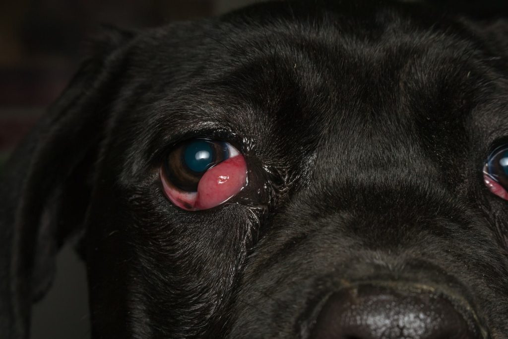
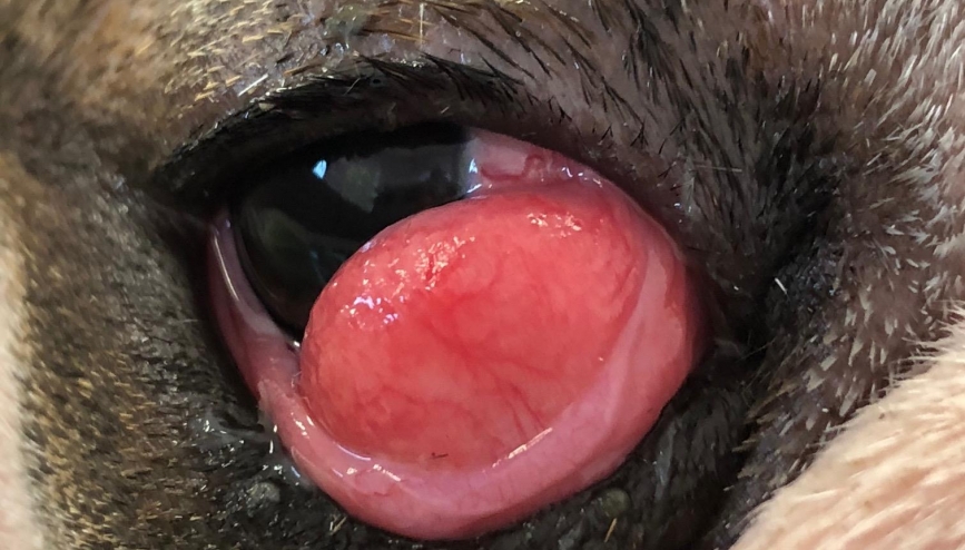
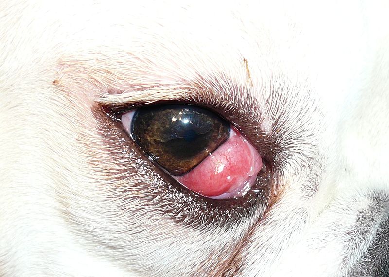
It occurs when the gland of the third eyelid (nictitating membrane) protrudes or prolapses from its normal position, resulting in the appearance of a reddish or pinkish mass resembling a cherry, hence the name.
This article aims to provide a comprehensive overview of cherry eye in animals, covering its disease entity, causes, clinical signs, diagnosis, treatment options, prognosis, and relevant references.
Cherry Eye Disease Entity
Cherry eye is characterized by the prolapse or protrusion of the gland of the third eyelid, also known as the nictitans gland or the gland of the third eyelid.
This gland plays a vital role in producing tears and maintaining ocular lubrication. When the gland prolapses, it becomes exposed and visible in the corner of the affected eye, presenting as a reddish or pinkish mass resembling a cherry.
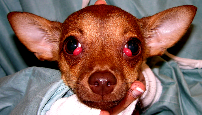
While cherry eye typically occurs unilaterally, it can affect both eyes in some cases.
Causes
The precise cause of cherry eye in animals is not fully understood, but several factors may contribute to its development.
These factors include:
- Weakness or laxity of the connective tissue that supports the nictitans gland.
- Breed predisposition, with certain breeds such as Bulldogs, Cocker Spaniels, and Beagles being more commonly affected.
- Trauma or injury to the eye or surrounding structures.
- Congenital predisposition or genetic factors.
While the underlying cause may vary among individual cases, cherry eye is generally considered a multifactorial condition with a genetic predisposition in certain breeds.
Cherry Eye Clinical Signs
The most prominent clinical sign of cherry eye in animals is the presence of a reddish or pinkish mass protruding from the corner of the affected eye, resembling a cherry. Other clinical signs may include:
- Ocular irritation or discomfort leads to rubbing or pawing at the affected eye.
- Increased tear production (epiphora) due to ocular irritation.
- Conjunctivitis or inflammation of the conjunctiva surrounding the prolapsed gland.
- Secondary ocular complications such as corneal ulceration or keratoconjunctivitis sicca (dry eye) if left untreated.
Diagnosis
Diagnosing cherry eye in animals is usually straightforward based on the characteristic appearance of the prolapsed gland.
However, additional diagnostic tests may be performed to assess ocular health and rule out underlying conditions.
Diagnostic steps may include:
- Ophthalmic examination: A thorough examination of the affected eye by a veterinarian or veterinary ophthalmologist to confirm the presence of cherry eye and evaluate ocular health.
- Fluorescein staining: Application of fluorescein dye to the eye to assess for corneal ulceration or other ocular surface abnormalities.
- Schirmer tear test: Measurement of tear production to evaluate for the presence of dry eye (keratoconjunctivitis sicca), which may occur secondary to cherry eye in some cases.
Cherry eye Treatment
The treatment of cherry eye in animals typically involves surgical intervention to reposition or replace the prolapsed gland and restore normal anatomy and function. Common treatment options include:
- Surgical replacement (tuck) technique: Surgical repositioning of the prolapsed gland using sutures or tissue adhesives to secure it in its normal anatomical position.
- Surgical removal (excision) technique: Complete or partial removal of the prolapsed gland, which may be considered in cases of severe or recurrent cherry eye.
Prognosis
The prognosis for animals with cherry eye is generally favorable following appropriate surgical treatment.
However, the risk of recurrence or development of complications such as dry eye (keratoconjunctivitis sicca) should be monitored postoperatively.
With prompt diagnosis and timely surgical intervention, most animals can achieve a successful outcome and return to normal ocular function.
HOW TO TAKE SLIT-LAMP EXAM IMAGES WITH A SMARTPHONE?
Smartphone slit-lamp photography is the new advancement in the field of science and technology in which photographs of the desired slit-lamp finding can be taken with smartphones by using the slit-lamp adapters.
Slit-lamp Smartphone photography
References
- Davidson, H. J., & Kuonen, V. J. (2005). Disorders of the canine third eyelid gland. Veterinary Clinics of North America: Small Animal Practice, 35(1), 213-232.
- Giuliano, E. A., Giuliano, A. M., & Moore, C. P. (2006). Treatment of cherry eye in dogs: a new minimally invasive technique. Veterinary Ophthalmology, 9(5), 291-296.
- Park, S. A., Ahn, H. J., Lee, S. H., & Yang, J. W. (2019). Clinical characteristics and surgical outcomes of cherry eye in 35 dogs (39 eyes). Veterinary Ophthalmology, 22(3), 354-360.
- Ramsey, D. T., & Plummer, C. E. (2013). Cherry eye. Clinical Techniques in Small Animal Practice, 28(2), 104-107.
