

CASE REPORT
DISEASE
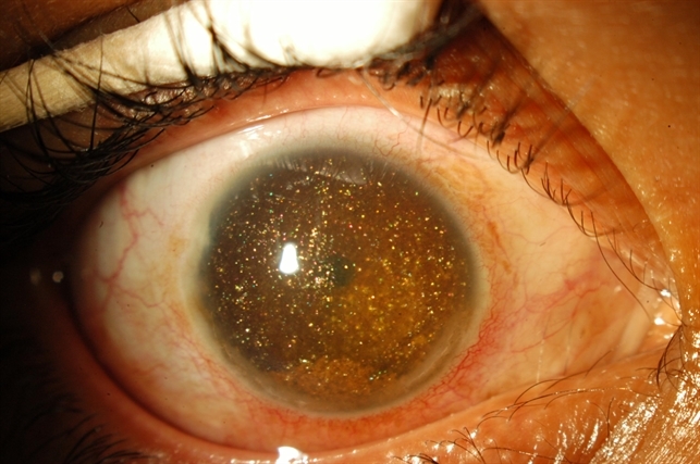
Synchysis scintillans, or cholesterolosis bulbi, is a rare degenerative ocular condition characterized by the accumulation of cholesterol crystals in the vitreous humor of the eye.

Cholesterol crystals appear as small, highly refractive opacities in the posterior chamber of the eye that freely move in a gravity-dependent manner, giving a snow globe-like effect.
There may also be anterior chamber involvement. While it is typically found in severely diseased eyes, synchysis scintillans is often an incidental asymptomatic finding.
The naming of the condition has been a subject of contention, most notably by Potter, who in 1983 argued that the pathological designation of “cholesterolosis bulbi” is more precise than the clinical descriptor “synchysis scintillans. It is often found incidentally.
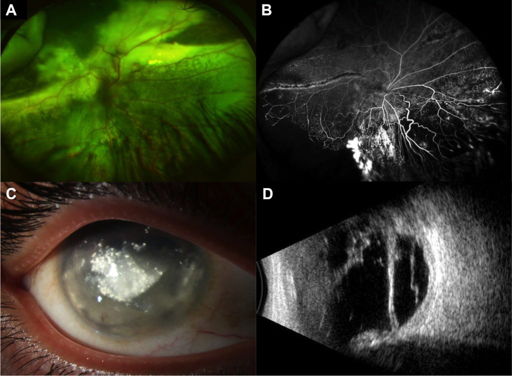
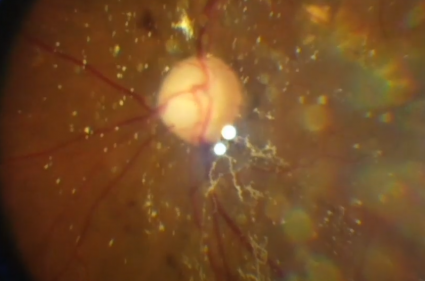
On fundoscopy, yellow, highly reflective, flat, angular crystals can be seen in the vitreous humor, which has been liquefied via syneresis. Liquefaction of the vitreous results in the opacities settling inferiorly in a gravity–dependent manner when the eye is immobilized.
Synchysis scintillans is postulated to be due to a degenerative process in the vitreous. This can happen in any age group due to other ocular conditions like vitreous hemorrhage, hyphema, retinal detachment, or chronic uveitis.
Rarely, when the vitreous gains access to the anterior chamber (e.g., in aphakia), synchysis scintillans of the anterior chamber can also be seen.
MANAGEMENT
Uveitis or vascular workup may be required to rule out active or lingering inflammation, which can produce vitritis.
Since vitreous hemorrhage is associated with degenerative processes that can raise intraocular pressure, patients should also be monitored for glaucoma.
Treatment
Since synchysis scintillans is considered a non-progressive and asymptomatic ocular condition, treatment is usually unnecessary.
However, the condition may be the result of underlying ocular diseases such as chronic uveitis, vitreous hemorrhage, retinal detachment, raised intraocular pressure, or inflammation. These underlying conditions should be treated accordingly.
A therapeutic strategy that targets oxidative stress may be effective in the treatment of synchysis patients. However, no pharmacological studies have been done to assess this strategy.
In the case of a patient with glaucoma and cholesterolosis bulbi in both the anterior and posterior segments, pars plana vitrectomy was performed. Not all cholesterol crystals could be removed due to a continuous influx of crystals.
While the vitrectomy led to a successful decrease in intraocular pressure and bevacizumab injection led to iris neovascularization regression, the number of cholesterol crystals did not decrease significantly.
Historically, anterior segment cholesterolosis has been treated with enucleation due to intractable pain and a risk of contralateral eye involvement leading to inflammation of sympathetic ophthalmia.
Would you have interest in taking retina images by smartphone?
Fundus photography is superior to fundus analysis as it enables intraocular pathologies to be photo captured and encrypted information to be shared with colleagues and patients.
Recent technologies allow smartphone-based attachments and integrated lens adaptors to transform the smartphone into a portable fundus camera and Retinal imaging by smartphone.
RETINAL IMAGING BY YOUR SMARTPHONE
REFERENCES
- Wand M, Smith TR, Cogan DG. Cholesterosis Bulbi: The Ocular Abnormality Known as Synchysis Scintillans. American Journal of Ophthalmology. 1975;80(2):177-183. doi:10.1016/0002-9394(75)90129-4
- Gonçalves M.B. (2020) Asteroid Hyalosis and Synchysis Scintillans. In: Rodrigues E., Meyer C, Tomazoni E. (eds) Trauma and Miscellaneous Disorders in Retina. Retina Atlas. Springer, Singapore. Doi:10.1007/978-981-13-8550-6
- Kumar S. Cholesterol crystals in the anterior chamber. Br J Ophthalmol 1963;47:295-9.
- Potter JW. Synchysis Scintillans, Cholesterosis Bulbi, and Asteroid Bodies: Does Synchysis Scintillans Exist? The Australian Journal of Optometry. 1983;66(6):232-238. doi:10.1111/j.1444-0938.1983.tb03724.
- Margo CE. Age-Related Diseases of the Vitreous. In: Cavallotti CAP, Cerulli L, eds. Age-Related Changes of the Human Eye. Aging Medicine. Humana Press; 2008:157-191. doi:10.1007/978-1-59745-507-7_8
RETINAL IMAGING BY YOUR SMARTPHONE

RETINAL IMAGING BY YOUR SMARTPHONE