
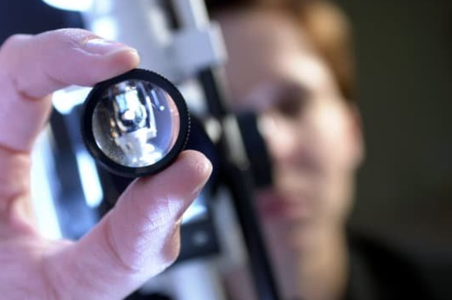
The Slit lamp examination is also known as biomicroscopy because it allows the in vivo study of living tissues of eye under the high magnification. is also known as biomicroscopy because it allows the in vivo study of living tissues of eye under the high magnification.
slit lamp consists of two parts, an illumination system and a low-power binocular microscope mounted horizontally.


Document what you see
Two smartphone imaging tools built for everyday clinic use — one for the slit lamp, one for the fundus.
From Choroida — the team behind this siteProcedure of Slit lamp Exam
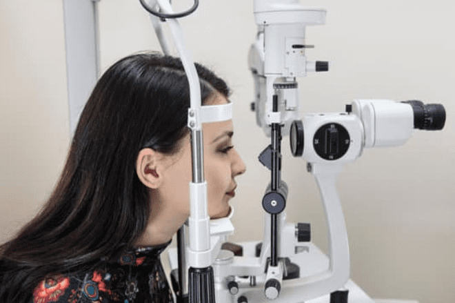
The examiner and patient are seated in either side of Slit lamp examination, the patient steadying his head by placing his chin on a chin rest and his forehead against a support.
The examiner usually views first the anterior segment. The fundus examination is carried out by interposing an additional lens such as Hruby lens (-55D), 90D or 78D lens.
Slit lamp photography of every part for documentation can be done with the help of slit lamp cameras and smartphones. Smartphones can be attached to slit lamps with the help of slit lamp adapters.
How to use Slit lamp examination for eye ?
There are some conditions, filters and illumination techniques for biomicroscopic examination.
- Biomicroscopic examination should be carried into a semi dark room.
- Both the patient and examiner should be positioned comfortably.
- Diffuse illumination should be used for as short a time as necessary.
- Adjust the illumination or brightness through the desired filters.
- Adjust the width and height of the beam.
- Low magnification should be first used to detect any pathology.
Methods of illumination in Slit lamp examination
1. Diffuse illumination:
In this technique
- Angle between microscope and illumination system should be 30-45°.
- Slit width should be widest.
- Magnification used is low to medium.
- Illumination should be medium to high.
It is used for
- General view of anterior eye and the palpebral conjunctiva
2. Direct focal illumination:
The slit beam is accurately focused upon that part of the eye under expection.
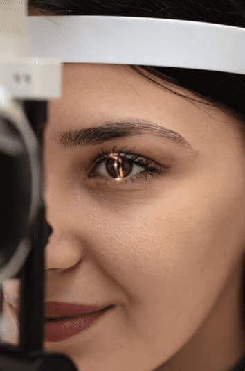
Light is directed as a narrow slit at an oblique angle (30-45°). Heterogenous tissues like cornea and lens disperse light and become visible as bright objects against a dark background.
3. Conical beam:
It is a small circular beam used to examine the presence of aqueous flare. beam is focused between the cornea and anterior lens surface and the dark zone between the cornea and the lens is observed. Flare appears grey or milky, seen as white dots. Settings of Slit lamp examination are
- Light source: 45-60° temporally and directed into the pupil
- Biomicroscope: directly in front of the eye
- Magnification: high (16-20x)
4. Indirect illumination:
- The slit beam is focused on a position just beside the area to be examined.
- Angle between slit lamp and microscope should be 30-45°.
- Beam width used is moderate.
It is useful to observe:
- Corneal infiltrates
- Corneal microcysts
- Epithelial cells.
5. Retro-illumination:
This technique involves illuminating the structure being examined reflected from a structure behind it. E.g. iris atrophy is identified by light which is reflected from the choroid.
Specular reflection
A great deal of information can be gained about the nature of a mirror-like surface by examining the rays of light reflected from it e.g. corneal surfaces and anterior lens capsules
Sclerotic scatter
It is used to outline even the faintest corneal pathology. Light beam is focused at the limbus. Because of the total internal reflection, rays of light pass through the substance of cornea and illuminate the opposite side of limbus.
Filters used in Slit lamp bio microscopy examination
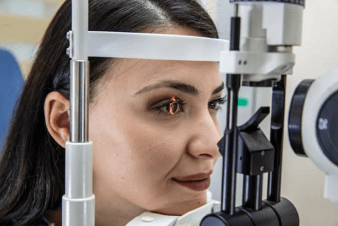
- Cobalt blue filter
It is used to check the tearfilm of the eye and in applanation tonometry.
- Green filter (red free filter)
It is used to view the blood vessels and hemorrhage
- Neutral density filter
It is used to decrease the maximum brightness for photosensitive patients
- Yellow filter
It is used for good contrast enhancement
- Diffuser filter
Used for general observations of the eye.
- Heat absorbing filters
It decreases patient discomfort.
Do you find this article useful? let’s know in a comment.. Thank you
Read about:
Test yourself
A few questions straight from this article.
-
Why is slit lamp examination also called biomicroscopy?
The slit lamp is termed a biomicroscope because it permits in vivo study of the living tissues of the eye under high magnification. -
Which two components make up a slit lamp?
A slit lamp consists of an illumination system together with a low-power binocular microscope mounted horizontally. -
Which lenses are interposed to examine the fundus at the slit lamp?
After the anterior segment is viewed, fundus examination is carried out by interposing an additional lens such as the Hruby (-55 D), 90 D or 78 D lens. -
Which magnification should be used first when starting a slit lamp examination?
Low magnification is used first to detect any pathology, and the examination is conducted in a semi-dark room with both patient and examiner comfortably positioned. -
Which settings define diffuse illumination at the slit lamp?
Diffuse illumination uses the widest slit width, a 30-45 degree angle between microscope and illumination, and low to medium magnification for a general view of the anterior eye. -
What is the conical beam technique at the slit lamp used to detect?
A small circular conical beam is focused in the dark zone between cornea and anterior lens surface; flare appears grey or milky and cells are seen as white dots. -
How is the beam positioned for indirect illumination at the slit lamp?
In indirect illumination the slit beam is focused just beside the area of interest, using a moderate beam width, which helps show corneal infiltrates, microcysts and epithelial cells. -
Which slit lamp technique outlines even the faintest corneal pathology?
In sclerotic scatter the beam is focused at the limbus and total internal reflection carries light through the corneal substance, illuminating the opposite limbus and outlining faint corneal pathology. -
What is the green (red-free) slit lamp filter used to view?
The green or red-free filter is used to view blood vessels and haemorrhage, whereas the cobalt blue filter serves for the tear film and applanation tonometry. -
Which slit lamp filter is used for tear film assessment and applanation tonometry?
The cobalt blue filter is used to check the tear film and during applanation tonometry, while the neutral density filter reduces brightness for photosensitive patients.
