LHEP (lamellar hole-associated epiretinal proliferation) is a thick epiretinal tissue found around many lamellar macular holes.
Pang, Spaide and Freund described it as a distinct entity in 2014. On OCT, however, it looks quite different from a classic epiretinal membrane, and therefore recognizing it changes how you classify the hole and plan surgery.
What LHEP Looks Like on OCT
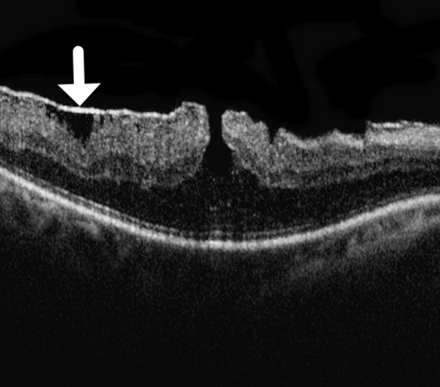
LHEP appears as a thick, homogeneous band of moderate reflectivity on the inner retinal surface. Its signal resembles the outer plexiform layer, while a tractional membrane is thin and bright. The tissue usually thickens toward the hole and merges with the middle retinal layers at the edge, and it lacks the folds and contraction typical of a macular pucker.
Clinical Appearance at the Slit Lamp
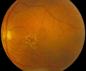
On biomicroscopy, the tissue may look like a yellowish or grayish film over the fovea, but the sign is subtle and easy to miss without OCT. Some patients report mild blur or reading difficulty, while others have no symptoms.
Fundus Explorer Pro
Photograph the retinal findings described here with the phone already in your pocket — 22 D optics and built-in illumination in one handheld unit.
From Choroida — the team behind this siteLHEP Versus Tractional ERM
Both membranes can coexist in one eye, so compare them feature by feature.
- Tractional ERM: thin, hyperreflective and contractile, with inner retinal wrinkling
- LHEP: thick and isoreflective with the middle layers, without visible contraction
- Tractional ERM: often creates schisis between the outer plexiform and outer nuclear layers, while the ellipsoid zone stays intact
- LHEP: covers a cavity with rounded, undermined edges and more frequent ellipsoid zone defects
Also, for a wider comparison of traction patterns, see ERM versus VMT on OCT.
Where LHEP Fits in the Consensus Definition
Hubschman and colleagues proposed an OCT-based definition of lamellar macular hole in 2020. Mandatory features are an irregular foveal contour, a cavity with undermined edges, and apparent foveal tissue loss. LHEP is one optional supporting feature, together with a central foveal bump and ellipsoid zone disruption.
Eyes with a tractional membrane and schisis but no tissue loss are classed as ERM foveoschisis. As a result, LHEP points toward a true lamellar hole (see lamellar versus full-thickness holes).
Degenerative and Tractional Lamellar Holes
Govetto and colleagues described two phenotypes in 2016, in which tractional holes show a contractile membrane, schisis and a mostly intact ellipsoid zone. Degenerative holes, by contrast, show LHEP, a rounded cavity and more frequent outer retinal defects. Histologic studies suggest a glial origin, which fits a reparative response to tissue loss.
LHEP and Visual Function
Eyes with LHEP tend to have deeper defects and thinner central retina. Pang and colleagues also reported poorer visual acuity compared with lamellar holes without this tissue. However, acuity varies widely, and ellipsoid zone integrity appears to guide functional prognosis.
Natural History of Degenerative Holes
Most degenerative lamellar holes change slowly, and some enlarge gradually, while a minority progress to a full-thickness hole. Therefore, document the baseline carefully and repeat OCT at fixed intervals, often every 6 to 12 months when stable.
Imaging Tips for LHEP
First, use dense raster scans through the fovea, because a single line can miss the edge. Next, add radial scans and en face images to judge the extent of the tissue. Finally, compare its reflectivity with the outer plexiform layer before you call it a membrane.
Deciding on Surgery
Surgery suits eyes with progressive visual loss, increasing distortion or documented enlargement, while stable eyes with good acuity usually do well with observation. The evidence comes mostly from retrospective series, so indications vary between surgeons, and you should discuss lens status because vitrectomy accelerates nuclear cataract in phakic eyes.
Handling LHEP During Vitrectomy
Complete peeling of the tissue may enlarge the defect and risk a full-thickness hole. Therefore, many surgeons dissect it from the periphery toward the hole and keep it attached at the edge. They then embed the tissue into the cavity, a technique reported by Shiraga and colleagues, while foveal-sparing ILM peeling and gas tamponade are used variably, without strong comparative evidence.
Expected Results After Surgery
Most series report a better foveal contour after embedding or careful dissection. Visual gains tend to be modest, especially when the ellipsoid zone is already damaged, and in addition a postoperative full-thickness hole remains a recognized complication.
Counseling Patients With LHEP
Explain that the tissue is a slow healing response, and it rarely pulls on the retina. Then describe the surgical goal as stabilization, with a limited chance of large gains, and teach monocular Amsler grid checks at home.
Pitfalls to Avoid
- Do not treat LHEP as a contractile ERM and peel it aggressively
- Never diagnose a lamellar hole from a single scan line
- Do not confuse ERM foveoschisis with a degenerative lamellar hole


Document what you see
Two smartphone imaging tools built for everyday clinic use — one for the slit lamp, one for the fundus.
From Choroida — the team behind this siteReferences
- Pang CE, Spaide RF, Freund KB. Epiretinal proliferation seen in association with lamellar macular holes: a distinct clinical entity. Retina. 2014;34:1513-1523.
- Pang CE, Spaide RF, Freund KB. Comparing functional and morphologic characteristics of lamellar macular holes with and without lamellar hole-associated epiretinal proliferation. Retina. 2015;35:720-726.
- Govetto A, Dacquay Y, Farajzadeh M, et al. Lamellar macular hole: two distinct clinical entities? Am J Ophthalmol. 2016;164:99-109.
- Hubschman JP, Govetto A, Spaide RF, et al. Optical coherence tomography-based consensus definition for lamellar macular hole. Br J Ophthalmol. 2020;104:1741-1747.
- Shiraga F, Takasu I, Fukuda K, et al. Modified vitreous surgery for symptomatic lamellar macular hole with epiretinal membrane containing macular pigment. Retina. 2013;33:1263-1269.
Test yourself
A few questions straight from this article.
-
How does lamellar hole-associated epiretinal proliferation (LHEP) appear on OCT?
LHEP is a thick, homogeneous band on the inner retinal surface whose signal resembles the outer plexiform layer, while a tractional membrane is thin and bright. -
Which feature distinguishes LHEP from a tractional epiretinal membrane on OCT?
Tractional ERM is thin, hyperreflective and contractile with inner retinal wrinkling, whereas LHEP is thick, isoreflective with the middle layers and shows no visible contraction. -
In the 2020 OCT-based consensus definition of lamellar macular hole, how is LHEP classified?
The mandatory features are an irregular foveal contour, a cavity with undermined edges and apparent foveal tissue loss; LHEP is optional, alongside a foveal bump and ellipsoid zone disruption. -
Under the lamellar macular hole consensus, an eye with a tractional membrane and schisis but no foveal tissue loss is classed as what?
Eyes with a tractional membrane and schisis but no tissue loss are classed as ERM foveoschisis, so the presence of LHEP points toward a true lamellar hole. -
Which lamellar macular hole phenotype described by Govetto and colleagues is associated with LHEP?
Degenerative holes show LHEP, a rounded cavity and more frequent outer retinal defects, whereas tractional holes show a contractile membrane, schisis and a mostly intact ellipsoid zone. -
What origin do histologic studies suggest for LHEP tissue?
Histologic studies suggest a glial origin, which fits LHEP being a reparative response to tissue loss. -
In eyes with LHEP, which OCT feature appears to guide functional prognosis?
Eyes with LHEP tend to have deeper defects and poorer acuity, but acuity varies widely and ellipsoid zone integrity appears to guide functional prognosis. -
How often is OCT typically repeated for a stable degenerative lamellar hole with LHEP?
Most degenerative holes change slowly, so the baseline is documented carefully and OCT is repeated at fixed intervals, often every 6 to 12 months when stable. -
Why do many surgeons avoid complete peeling of LHEP during vitrectomy?
Complete peeling may enlarge the defect and risk a full-thickness hole, so many surgeons dissect it toward the hole, keep it attached and embed it into the cavity. -
Why should lens status be discussed before vitrectomy for a lamellar hole with LHEP?
Surgery suits progressive or symptomatic eyes, and lens status matters because vitrectomy accelerates nuclear cataract in phakic eyes.

Writes and reviews the clinical articles on Educate. Every post is checked against peer-reviewed sources before it goes live.