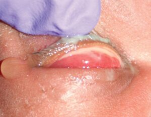
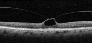
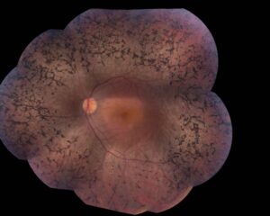
Trachoma grading uses the WHO simplified system to record five signs of ocular chlamydial disease.
The examiner needs only 2.5x loupes, good light and an everted upper lid. Each sign is graded separately, so one eye can carry several grades at once.
Origins of Trachoma Grading
Thylefors and colleagues published the simplified system in 1987 for trained nonspecialist graders, and it replaced detailed research scales that required slit lamps and expert observers. In 2020, a WHO working group amended the definitions of TT and CO. As a result, current surveys grade trichiasis on the upper lid only.
Preparing the Lid for Trachoma Grading
First, inspect the lashes and cornea before touching the lid, then evert the upper lid and expose the tarsal plate (see eyelid eversion technique). Grade the central tarsal conjunctiva and ignore the edges and fornix.
All-fit Slit-Lamp Adapter
Record and share exactly what you see at the slit lamp. One adapter fits any slit lamp or surgical microscope — and any smartphone.
From Choroida — the team behind this siteTF: Trachomatous Inflammation, Follicular
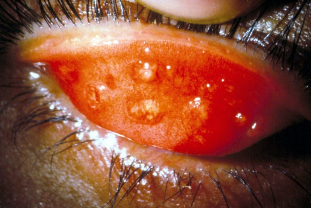
TF requires five or more follicles in the central upper tarsal conjunctiva, and each follicle must measure at least 0.5 mm across. Follicles appear as pale, round, slightly gelatinous elevations, but follicles at the tarsal corners or in the fornix do not count.
Why TF Drives Trachoma Grading Surveys
TF is most common in young children, who carry the largest chlamydial load. Therefore, population surveys measure TF prevalence in children aged 1 to 9 years, and district prevalence then decides whether mass drug administration is needed.
TI: Trachomatous Inflammation, Intense
TI means pronounced inflammatory thickening of the tarsal conjunctiva, which obscures more than half of the normal deep tarsal vessels. These vessels run vertically and are usually easy to see, but papillary hypertrophy and edema hide them, so the surface looks red and velvety. In addition, TI often coexists with TF and signals more severe infection.
TS: Trachomatous Scarring
TS describes easily visible white lines, bands or sheets in the tarsal conjunctiva, and a horizontal scar near the lid margin forms the classic Arlt’s line. Scarring reflects years of repeated infection and inflammation. However, TS alone needs no antibiotics, while it does need watching for evolving trichiasis.
TT: Trachomatous Trichiasis
TT is present when at least one upper lid lash touches the eyeball, and evidence of recent removal of in-turned upper lid lashes also counts. Therefore, look for broken lash stubs, because many patients epilate at home.
Separating TT From Other Lash Problems in Trachoma Grading
A scarred, shortened tarsus with entropion supports a trachomatous cause, but confirm TS on the same lid before blaming trachoma. Without scarring, consider blepharitis, trauma or cicatricial pemphigoid (see trichiasis).
CO: Corneal Opacity
CO is an easily visible corneal opacity over the pupil, and under the amended definition, the opacity blurs at least part of the pupil margin. Peripheral scars away from the visual axis do not meet this grade. As a result, CO captures the opacities most likely to reduce vision.
Herbert’s Pits and Signs Outside Trachoma Grading
Herbert’s pits are limbal depressions left by healed follicles, and they support the diagnosis but carry no grade in the simplified system. Similarly, pannus and limbal follicles are not graded, although clinic notes can record them (see trachoma).
Mapping Trachoma Grading to Antibiotic Treatment
The SAFE strategy combines surgery, antibiotics, facial cleanliness and environmental improvement, and TF prevalence guides the antibiotic arm at district level. Districts above the threshold receive mass azithromycin, with the number of annual rounds set by baseline prevalence. Individual patients with TF or TI also receive azithromycin or topical tetracycline ointment.
Mapping TT to Trichiasis Surgery
TT is the grade that triggers referral for lid surgery, and bilamellar and posterior lamellar tarsal rotation are the main procedures. In an Ethiopian randomized trial, posterior lamellar rotation produced less postoperative trichiasis. However, recurrence still occurs with either technique, so follow-up matters.
Counseling Patients With CO
CO marks damage that antibiotics and lid surgery cannot reverse, so surgery protects the remaining clear cornea while existing scars persist. Explain this before the operation to keep expectations realistic. Keratoplasty has a limited role, because the ocular surface is dry and scarred.
Elimination Targets and Trachoma Grading
WHO uses two prevalence targets to declare elimination as a public health problem. First, TF must fall below 5% in children aged 1 to 9 years, and next, TT unknown to the health system must fall below 0.2% in adults aged 15 and older. Accurate grading at both ends of the disease is therefore essential.
Pitfalls to Avoid
- Counting follicles at the tarsal edge or fornix as TF
- Grading TI from redness alone without checking deep vessels
- Missing TT because the patient recently epilated
- Recording lower lid trichiasis as TT under the 2020 definition
- Calling peripheral corneal scars CO


Document what you see
Two smartphone imaging tools built for everyday clinic use — one for the slit lamp, one for the fundus.
From Choroida — the team behind this siteReferences
- Thylefors B, Dawson CR, Jones BR, West SK, Taylor HR. A simple system for the assessment of trachoma and its complications. Bull World Health Organ. 1987;65:477-483.
- Solomon AW, Kello AB, Bangert M, et al. The simplified trachoma grading system, amended. Bull World Health Organ. 2020;98:698-705.
- Taylor HR, Burton MJ, Haddad D, West S, Wright H. Trachoma. Lancet. 2014;384:2142-2152.
- Habtamu E, Wondie T, Aweke S, et al. Posterior lamellar versus bilamellar tarsal rotation surgery for trachomatous trichiasis in Ethiopia: a randomised controlled trial. Lancet Glob Health. 2016;4:e175-e184.
Test yourself
A few questions straight from this article.
-
What defines TF (trachomatous inflammation, follicular) in the WHO simplified trachoma grading?
TF requires five or more follicles, each at least 0.5 mm, in the central upper tarsal conjunctiva; follicles at the tarsal corners or fornix do not count. -
In which age group do trachoma population surveys measure TF prevalence?
TF is most common in young children, who carry the largest chlamydial load, so surveys measure it in children aged 1 to 9; district prevalence then decides on mass drug administration. -
What defines TI (trachomatous inflammation, intense) in WHO trachoma grading?
TI is pronounced inflammatory thickening that hides more than half of the normally visible vertical deep tarsal vessels, so it should not be graded from redness alone. -
What does trachomatous scarring (TS) alone require?
TS reflects years of repeated infection; it needs no antibiotics on its own but should be watched for evolving trichiasis. -
What qualifies as TT (trachomatous trichiasis) in WHO trachoma grading?
TT is present when at least one upper lid lash touches the eyeball or there is evidence of recent removal of in-turned upper lid lashes, so look for broken stubs. -
Before attributing trichiasis to trachoma, which sign should be confirmed on the same lid?
A scarred, shortened tarsus with entropion supports a trachomatous cause, but TS should be confirmed on the same lid; without scarring, consider blepharitis, trauma or cicatricial pemphigoid. -
Under the amended WHO definition, which corneal opacity qualifies as CO in trachoma grading?
CO is an easily visible opacity over the pupil that blurs at least part of the pupil margin; peripheral scars away from the visual axis do not qualify. -
Which trichiasis operation produced less postoperative trichiasis in an Ethiopian randomized trial?
TT triggers referral for lid surgery; posterior lamellar rotation produced less postoperative trichiasis than bilamellar rotation, though recurrence occurs with either. -
What WHO elimination target applies to trachomatous trichiasis unknown to the health system?
Elimination as a public health problem requires TF below 5% in children aged 1 to 9 and TT unknown to the health system below 0.2% in adults aged 15 and older. -
What are Herbert's pits, and how are they handled in WHO simplified trachoma grading?
Herbert's pits are limbal depressions left by healed follicles; they support the diagnosis but carry no grade in the simplified system.

Writes and reviews the clinical articles on Educate. Every post is checked against peer-reviewed sources before it goes live.