Chorioretinal atrophy is loss of the retinal pigment epithelium, outer retina and choriocapillaris that exposes the sclera on fundus examination.
The white patches look alike across many diseases, but their pattern, location and the patient’s age usually narrow the cause quickly.
What Chorioretinal Atrophy Looks Like
The lesion appears white or yellow-white, with sharp or scalloped borders, and large choroidal vessels often cross the base when the choriocapillaris is gone but deeper vessels survive. Pigment clumps also commonly outline the edge, but when the whole choroid is lost, bare sclera shows with no visible vessels.
Sorting Chorioretinal Atrophy by Pattern
First, ask whether the atrophy is diffuse, patchy, geographic or linear, and next note where it sits: peripapillary, macular, midperipheral or far peripheral. Then check symmetry between the two eyes: bilateral symmetric disease suggests a genetic or degenerative cause, while unilateral lesions suggest trauma, infection or laser.
Fundus Explorer Pro
Photograph the retinal findings described here with the phone already in your pocket — 22 D optics and built-in illumination in one handheld unit.
From Choroida — the team behind this sitePathologic Myopia
Myopic maculopathy is the commonest cause in many clinics, and the META-PM system grades it from tessellated fundus to diffuse atrophy, patchy atrophy and macular atrophy. Lacquer cracks, a Fuchs spot and a posterior staphyloma also support the diagnosis, so check the refraction and axial length before ordering any genetic workup.
Myopic Chorioretinal Atrophy and CNV
Patchy atrophy can enlarge around a regressed choroidal neovascular membrane, and because new CNV still threatens vision, fresh hemorrhage or metamorphopsia needs OCT and OCT angiography. Anti-VEGF injections treat active CNV, but they cannot reverse established atrophy, and patients should hear that clearly.
Gyrate Atrophy
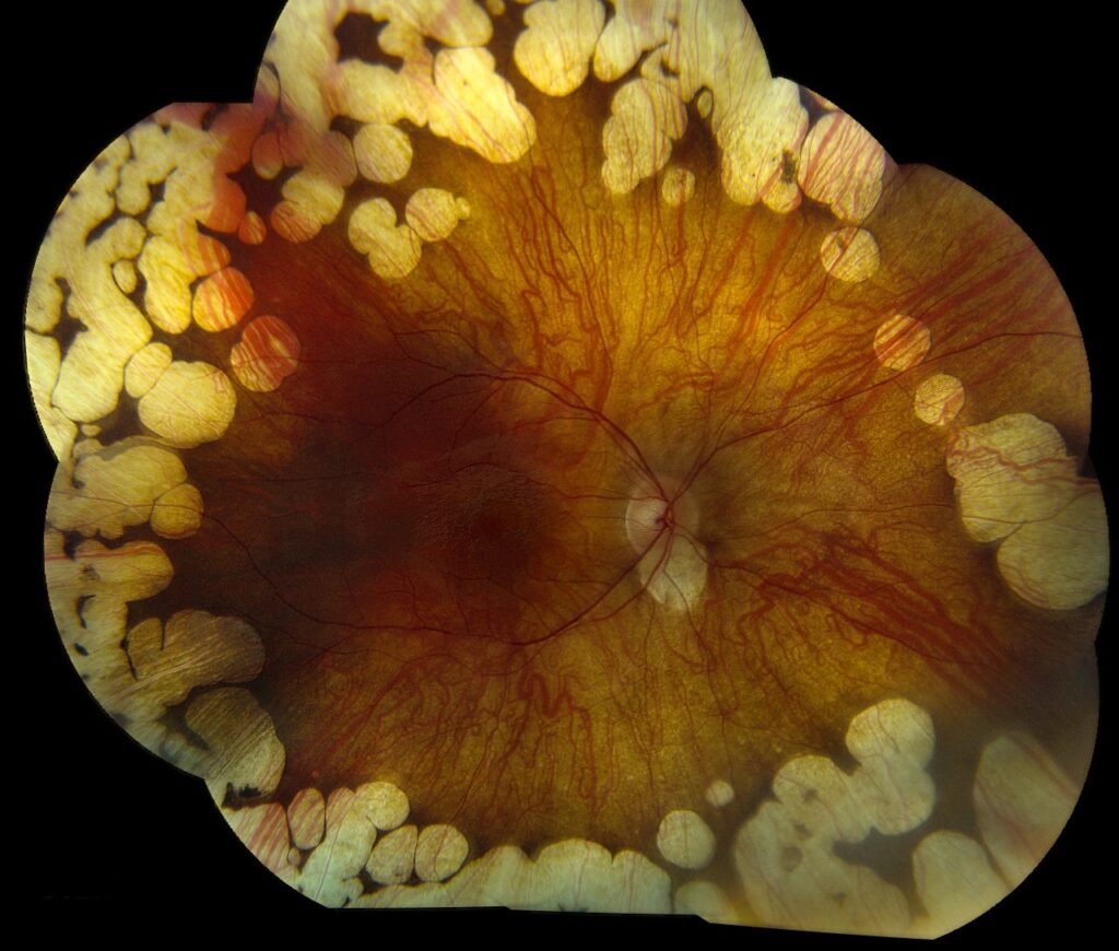
Gyrate atrophy causes sharply scalloped patches that start in the midperiphery and coalesce toward the posterior pole, and it results from ornithine aminotransferase deficiency, an autosomal recessive disorder. Patients present in childhood or adolescence with night blindness, high myopia and early cataract, and plasma ornithine is markedly raised, which confirms the diagnosis (see gyrate atrophy).
Treatment Limits in Gyrate Chorioretinal Atrophy
An arginine-restricted diet lowers ornithine and may slow progression in compliant patients, and a small subset responds to pyridoxine. However, diet adherence is hard, and the evidence comes mainly from small series, while cystoid macular edema also occurs and deserves separate treatment.
Choroideremia
Choroideremia is an X-linked disease caused by mutations in the CHM gene, and affected males report night blindness in the first decade and progressive peripheral field loss. The atrophy spreads centripetally, while a central island of macula survives until late adulthood.
Carrier females often show patchy peripheral pigment mottling (see choroideremia), and subretinal AAV gene therapy proved feasible in early trials. However, a phase 3 trial missed its primary endpoint, so treatment remains investigational.
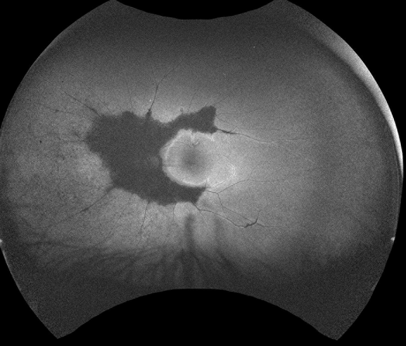
Inflammatory Chorioretinal Atrophy
Serpiginous choroiditis produces geographic atrophy that spreads from the disc in a snake-like fashion, and active edges look creamy and gray, and they stain on fluorescein angiography. Because tuberculosis can mimic it, test for latent TB before starting immunosuppression, and multifocal choroiditis and presumed ocular histoplasmosis also leave punched-out scars with peripapillary atrophy.
Traumatic Chorioretinal Atrophy
Chorioretinitis sclopetaria follows a high-velocity projectile passing near the globe without perforating it. The choroid and retina rupture, then heal with a white claw-like area of bare sclera.
In contrast, a choroidal rupture after blunt trauma forms a crescent concentric to the optic disc. Both lesions are unilateral, and the history usually explains them.
Laser Scars
Panretinal photocoagulation leaves round pigmented scars in regular rows, and these scars can enlarge over the years, so a lesion near the fovea may threaten vision later. Retinopexy around a break also leaves a ring of atrophy (see laser retinopexy). Therefore, regular spacing and a documented treatment history make the diagnosis simple.
Chorioretinal Atrophy by Age
Age gives a useful first filter.
- Childhood and adolescence: gyrate atrophy, choroideremia, congenital toxoplasmosis
- Young adults: myopic maculopathy, multifocal choroiditis, trauma
- Middle age: serpiginous choroiditis, histoplasmosis scars, laser scars
- Older adults: geographic atrophy in AMD, pavingstone degeneration, advanced myopia
Benign Peripheral Lookalikes
Pavingstone degeneration shows discrete round patches in the inferior periphery between the ora and equator, and large choroidal vessels are visible through the base. It is also common in older adults and needs no treatment, so recognize it to avoid needless referral.
Imaging Chorioretinal Atrophy
Fundus autofluorescence shows dark hypoautofluorescent areas where the RPE has died, and OCT shows outer retinal loss with hypertransmission into the choroid. In addition, full-field ERG separates widespread dystrophies from focal scars, and finally, plasma ornithine, genetic panels and infection serology confirm specific causes.
Pitfalls to Avoid
- Do not label every white patch as myopic before checking the refraction
- Never start steroids for serpiginous-like disease without excluding tuberculosis and syphilis
- Do not miss gyrate atrophy in a myopic child with night blindness
- Remember that drugs such as thioridazine can cause nummular atrophy


Document what you see
Two smartphone imaging tools built for everyday clinic use — one for the slit lamp, one for the fundus.
From Choroida — the team behind this siteReferences
- Ohno-Matsui K, Kawasaki R, Jonas JB, et al. International photographic classification and grading system for myopic maculopathy. Am J Ophthalmol. 2015;159:877-883.
- Takki KK, Milton RC. The natural history of gyrate atrophy of the choroid and retina. Ophthalmology. 1981;88:292-301.
- MacLaren RE, Groppe M, Barnard AR, et al. Retinal gene therapy in patients with choroideremia: initial findings from a phase 1/2 clinical trial. Lancet. 2014;383:1129-1137.
- Salmon JF. Kanski’s Clinical Ophthalmology: A Systematic Approach. 9th ed. Elsevier; 2020.
- Schachat AP, Sadda SR, Hinton DR, et al, eds. Ryan’s Retina. 6th ed. Elsevier; 2018.
Test yourself
A few questions straight from this article.
-
Which tissues are lost in chorioretinal atrophy, exposing the sclera on fundus examination?
Chorioretinal atrophy is loss of the RPE, outer retina and choriocapillaris; large choroidal vessels cross the base when deeper vessels survive, and bare sclera shows when the whole choroid is lost. -
Unilateral chorioretinal atrophy most suggests which group of causes?
Bilateral symmetric atrophy suggests a genetic or degenerative cause, whereas unilateral lesions suggest trauma, infection or laser. -
Which system grades myopic maculopathy from tessellated fundus through diffuse and patchy atrophy to macular atrophy?
META-PM grades myopic maculopathy from tessellated fundus to diffuse, patchy and macular atrophy; lacquer cracks, a Fuchs spot and staphyloma also support the diagnosis. -
What can anti-VEGF injections achieve in myopic chorioretinal atrophy complicated by choroidal neovascularization?
Anti-VEGF treats active CNV but cannot reverse established atrophy, which can enlarge around a regressed membrane — patients should be told this clearly. -
Which laboratory finding confirms gyrate atrophy of the choroid and retina?
Gyrate atrophy results from autosomal recessive ornithine aminotransferase deficiency, and markedly raised plasma ornithine confirms the diagnosis. -
Which dietary measure lowers ornithine and may slow progression of gyrate atrophy?
An arginine-restricted diet lowers ornithine and may slow progression in compliant patients, and a small subset responds to pyridoxine, though adherence is hard. -
How does the atrophy of choroideremia progress?
In X-linked choroideremia (CHM gene) the atrophy spreads centripetally while a central island of macula survives until late adulthood. -
What must be tested for before starting immunosuppression for serpiginous choroiditis?
Serpiginous choroiditis spreads from the disc in a snake-like fashion, and because tuberculosis can mimic it, latent TB should be tested for before immunosuppression. -
What shape does a choroidal rupture after blunt trauma typically form?
Blunt-trauma choroidal rupture forms a crescent concentric to the disc, whereas chorioretinitis sclopetaria from a near-miss projectile heals with a claw-like area of bare sclera. -
Where does pavingstone degeneration typically appear, and what does it need?
Pavingstone degeneration shows discrete round patches in the inferior periphery between the ora and equator, is common in older adults and needs no treatment.

Writes and reviews the clinical articles on Educate. Every post is checked against peer-reviewed sources before it goes live.