Interdigitation zone is the OCT term for the faint hyperreflective band between the ellipsoid zone and the retinal pigment epithelium.
It marks the contact between cone outer segment tips and the apical processes of the RPE. Because the band is thin and fragile, it often fades before other outer retinal bands, so careful reading can reveal early photoreceptor injury.
Anatomy of the Interdigitation Zone
The IN•OCT consensus panel named this band in 2014, and earlier papers called it the cone outer segment tip line, or COST line. Histologic models link it to the contact cylinder, where RPE microvilli wrap the outer segment tips. Rod outer segments also interdigitate with the RPE, although their share of the signal is less clear.
Band 3 or Band 4?
Numbering causes confusion in reports and in search results. In the common four band model, the external limiting membrane is band 1, the ellipsoid zone is band 2, the interdigitation zone is band 3, and the RPE and Bruch membrane complex forms band 4. However, some authors count differently, so name the band instead of quoting a number.
Fundus Explorer Pro
Photograph the retinal findings described here with the phone already in your pocket — 22 D optics and built-in illumination in one handheld unit.
From Choroida — the team behind this siteNormal Appearance on OCT
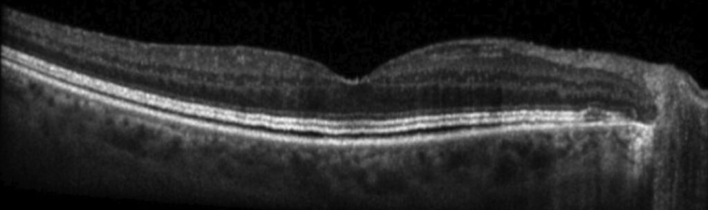
In a healthy fovea, the band runs parallel to the RPE and dips slightly beneath the foveal center, and its reflectivity is lower than the ellipsoid zone. It may merge with the RPE in older eyes or on lower resolution devices, so judge it only on averaged, well centered B-scans.
Why the Interdigitation Zone Is Fragile
Outer segments renew constantly and depend on close contact with healthy RPE, so when that contact loosens, the tips shorten or misalign first. As a result, the band can fade while the inner segment ellipsoids still reflect normally, and its signal depends on photoreceptor orientation, so beam tilt alone can weaken it.
Interdigitation Zone Loss Before Ellipsoid Zone Loss
Many outer retinal disorders follow a sequence in which the IZ fades, then the EZ breaks, then the ELM fails. This order is a useful clinical pattern, although it is not universal.
Once the ellipsoid zone breaks, the injury is usually more advanced (see ellipsoid zone disruption). Because of that sequence, an isolated IZ gap suggests early and possibly reversible damage.
Artifacts That Mimic Loss
- Oblique beam entry through a decentered pupil
- Media opacity and low signal strength
- Poor fixation or motion during the scan
- A scan line that misses the foveal center
Interdigitation Zone in MEWDS
Multiple evanescent white dot syndrome shows patchy IZ and EZ disruption around the disc and macula, and patients report photopsia and an enlarged blind spot. The white dots may fade before the visit, while OCT still shows outer band loss. Then, over several weeks, the bands usually reconstitute, and the IZ may return last.
Interdigitation Zone in AZOOR
Acute zonal occult outer retinopathy damages photoreceptors in zones that often begin at the disc, and the fundus may look normal early, so OCT carries much of the diagnosis. Typically, the IZ and EZ disappear within the zone, with a gradual transition at its border, and autofluorescence then outlines the trizonal pattern (see AZOOR).
Hydroxychloroquine Toxicity
Hydroxychloroquine retinopathy injures the parafoveal or pericentral outer retina, and in early cases the IZ may attenuate before the EZ breaks. Therefore, inspect the parafoveal ring carefully on every screening scan, while the AAO recommendations pair SD-OCT with automated visual fields for screening.
Inherited Dystrophies
In retinitis pigmentosa, the IZ usually ends closer to the fovea than the EZ edge, and as a result the band offers a sensitive marker of the transition zone. Cone dystrophies, by contrast, may show central IZ loss with better preserved bands outside the macula.
After Surgery and Detachment
After macular hole closure or detachment repair, outer bands often recover in stages, and in many series the ELM recovers first, then the EZ, then the IZ. Several reports link IZ restoration to better final acuity, but recovery may take months, so one early scan should not set the prognosis.
Grading the Interdigitation Zone
First, describe the band as present, attenuated or absent, and next, state its extent and location relative to the fovea. Compare the same scan position across visits using follow-up mode, and en face slabs can then map the loss more completely (see en face OCT).
Pitfalls to Avoid
- Do not call the band absent on a tilted scan
- Never rely on band numbers alone in reports
- Avoid judging recovery from one early scan
- Remember that a normal fundus does not exclude outer retinal disease


Document what you see
Two smartphone imaging tools built for everyday clinic use — one for the slit lamp, one for the fundus.
From Choroida — the team behind this siteReferences
- Staurenghi G, Sadda S, Chakravarthy U, Spaide RF. Proposed lexicon for anatomic landmarks in normal posterior segment spectral-domain optical coherence tomography: the IN•OCT consensus. Ophthalmology. 2014;121:1572-1578.
- Spaide RF, Curcio CA. Anatomical correlates to the bands seen in the outer retina by optical coherence tomography: literature review and model. Retina. 2011;31:1609-1619.
- Marmor MF, Kellner U, Lai TY, Melles RB, Mieler WF. Recommendations on screening for chloroquine and hydroxychloroquine retinopathy (2016 revision). Ophthalmology. 2016;123:1386-1394.
- Mrejen S, Khan S, Gallego-Pinazo R, Jampol LM, Yannuzzi LA. Acute zonal occult outer retinopathy: a classification based on multimodal imaging. JAMA Ophthalmol. 2014;132:1089-1098.
- Spaide RF, Koizumi H, Freund KB. Photoreceptor outer segment abnormalities as a cause of blind spot enlargement in acute zonal occult outer retinopathy-complex diseases. Am J Ophthalmol. 2008;146:111-120.
Test yourself
A few questions straight from this article.
-
What anatomical contact does the interdigitation zone on OCT represent?
The interdigitation zone marks where cone outer segment tips contact the apical processes of the RPE; histologic models link it to the contact cylinder where RPE microvilli wrap the tips. -
Why should an OCT report name the interdigitation zone rather than quote its band number?
In the common four-band model the interdigitation zone is band 3, but some authors count differently, so naming the band avoids confusion. -
How does a normal interdigitation zone appear at the healthy fovea on OCT?
In a healthy fovea the band runs parallel to the RPE, dips slightly beneath the foveal center and is less reflective than the ellipsoid zone. -
In many outer retinal disorders, in what order are the outer OCT bands usually lost?
A common, though not universal, pattern is that the IZ fades first, then the EZ breaks, then the ELM fails. -
What does an isolated interdigitation zone gap with an intact ellipsoid zone suggest on OCT?
Because the IZ usually fades before the EZ breaks, an isolated IZ gap suggests early and possibly reversible damage; an EZ break usually means more advanced injury. -
Why can beam tilt alone weaken the interdigitation zone signal on OCT?
The band's signal depends on photoreceptor orientation, so oblique beam entry through a decentered pupil can mimic loss; it should not be called absent on a tilted scan. -
In multiple evanescent white dot syndrome, what usually happens to the disrupted interdigitation and ellipsoid zones?
MEWDS shows patchy IZ and EZ disruption around the disc and macula that usually reconstitutes over several weeks, with the IZ possibly returning last. -
After macular hole closure or detachment repair, in what order do the outer OCT bands usually recover?
In many series the ELM recovers first, then the EZ, then the IZ; recovery may take months, so one early scan should not set the prognosis. -
Where should the interdigitation zone be inspected most carefully on hydroxychloroquine screening OCT scans?
Hydroxychloroquine injures the parafoveal or pericentral outer retina, where the IZ may attenuate before the EZ breaks in early cases. -
In retinitis pigmentosa, why is the interdigitation zone a sensitive marker of the transition zone?
In retinitis pigmentosa the IZ usually ends closer to the fovea than the EZ edge, so it marks the transition zone sensitively; cone dystrophies instead may show central IZ loss.

Writes and reviews the clinical articles on Educate. Every post is checked against peer-reviewed sources before it goes live.