Keratitis types are grouped by cause, and the cause decides the treatment.
Infection, immune reactions, exposure, nerve loss and drug toxicity can all inflame the cornea, but each group leaves a distinct footprint at the slit lamp. Therefore, a careful look often narrows the diagnosis before any culture returns.
How Keratitis Types Are Classified
Clinicians first separate infectious from noninfectious disease, and next they note the depth: epithelial, stromal or endothelial. Location also helps, because central and peripheral lesions have different usual causes. Finally, the staining pattern points toward a mechanism.
Bacterial Keratitis
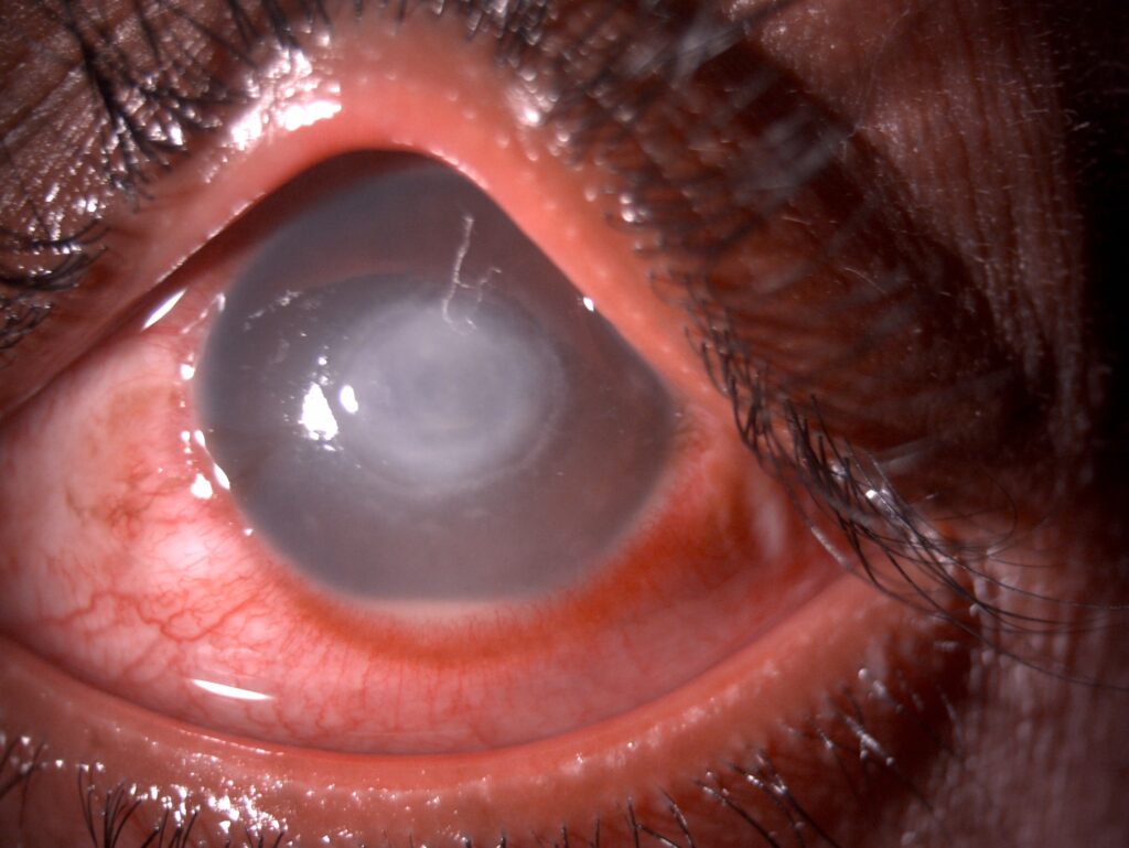
A bacterial ulcer shows an epithelial defect over a dense, white stromal infiltrate, and pain, discharge and anterior chamber reaction usually accompany it. In contact lens wearers, Pseudomonas can melt the stroma within a day (see Pseudomonas keratitis). As a result, this type demands same-day treatment.
All-fit Slit-Lamp Adapter
Record and share exactly what you see at the slit lamp. One adapter fits any slit lamp or surgical microscope — and any smartphone.
From Choroida — the team behind this siteFungal and Acanthamoeba Keratitis Types
Filamentous fungal ulcers have feathery margins, satellite lesions and a dry, raised surface, and hypopyon and endothelial plaque may appear despite a modest epithelial defect. In contrast, early Acanthamoeba shows a dirty epithelium and radial perineuritis. Later, a ring infiltrate may form, often with pain out of proportion to signs (see Acanthamoeba keratitis).
Herpes Simplex: Dendritic and Disciform Keratitis
Herpetic epithelial disease forms a branching dendrite with terminal bulbs that stains with fluorescein. Disciform keratitis, by contrast, presents as central stromal edema with an intact epithelium. Keratic precipitates sit beneath the edematous disc, and reduced corneal sensation also supports a herpetic cause.
Immune Keratitis Types at the Limbus
Marginal keratitis produces peripheral infiltrates with a clear zone next to the limbus and reflects hypersensitivity to staphylococcal antigens, usually with blepharitis. Peripheral ulcerative keratitis differs, because it causes stromal thinning at the limbus. Therefore, peripheral melting should prompt a search for systemic vasculitis.
Exposure Keratitis
Incomplete lid closure dries the inferior cornea first, so punctate staining in the lower third is the typical pattern. Facial nerve palsy, proptosis and lid scarring are common causes.
Neurotrophic Keratitis
Loss of corneal sensation impairs epithelial healing, and the defect is often oval and central, with smooth, rolled edges. Because patients report little pain, presentation is often late (see neurotrophic keratitis staging).
Toxic Keratitis
Preserved drops, topical anesthetic abuse and some antivirals injure the epithelium, and the staining is often inferonasal, where drops pool. As a result, stopping the offending drop is both diagnostic and therapeutic.
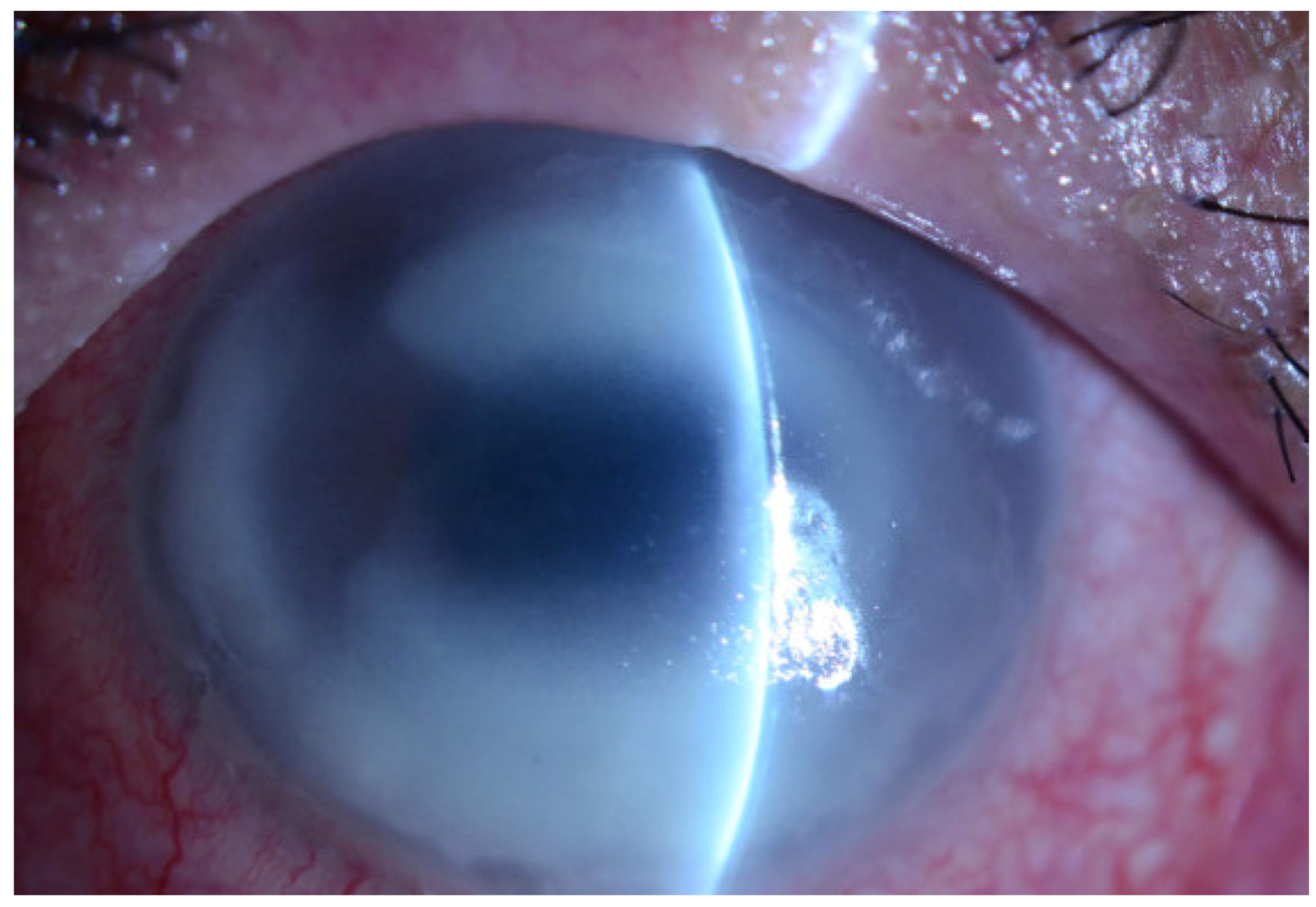
Filamentary Keratitis
Strands of mucus and epithelium attach to the cornea at one end, stain with lissamine green and cause foreign body sensation. Dry eye and prolonged patching are common associations. Therefore, treat the surface disease while removing the filaments.
Slit-Lamp Clues by Keratitis Types
- Bacterial: dense infiltrate under an epithelial defect
- Fungal: feathery edges and satellite lesions
- Acanthamoeba: radial perineuritis, then ring infiltrate
- Herpetic: dendrite with terminal bulbs
- Disciform: central stromal edema with keratic precipitates
- Marginal: peripheral infiltrate with a clear zone
- Exposure: inferior punctate staining
- Neurotrophic: oval defect with rolled edges
- Toxic: inferonasal staining after heavy drop use
- Filamentary: stained strands anchored to epithelium
When to Scrape in Keratitis Types
Culture large, central or deep ulcers before starting antibiotics, and also scrape atypical lesions and suspected fungal or Acanthamoeba cases. Small peripheral infiltrates can often be treated empirically. However, any worsening should trigger sampling.
Treating Keratitis Types by Cause
Bacterial ulcers need intensive broad-spectrum topical antibiotics, usually hourly at first. For filamentous fungal ulcers, MUTT found natamycin outperformed voriconazole, especially for Fusarium. Herpetic epithelial disease responds to topical or oral antivirals, while exposure, neurotrophic and toxic types need surface protection and removal of the cause.
The Steroid Question
Steroids help some types and harm others, and in the Herpetic Eye Disease Study, topical steroids with antiviral cover reduced stromal keratitis progression. SCUT, however, found no overall benefit of adjunctive steroids in bacterial ulcers. Because steroids worsen fungal and active epithelial herpetic disease, confirm the type first.
Counseling Patients with Keratitis Types
Explain that treatment depends on the cause, which may take days to confirm. Contact lens wearers must stop lens wear and bring the lenses and case. Finally, ask patients to return urgently with rising pain, discharge or worse vision.
Follow-Up in Keratitis Types
Review severe ulcers daily until the infiltrate stabilizes, and measure defect and infiltrate size each visit, because trends guide therapy. Then taper drops slowly once the epithelium heals.
Pitfalls to Avoid
- Do not start steroids before excluding fungus and active herpes
- Always test sensation before calling a painless defect healed
- Do not exclude Acanthamoeba on a negative culture
- Never treat peripheral melting as simple marginal keratitis


Document what you see
Two smartphone imaging tools built for everyday clinic use — one for the slit lamp, one for the fundus.
From Choroida — the team behind this siteReferences
- Austin A, Lietman T, Rose-Nussbaumer J. Update on the management of infectious keratitis. Ophthalmology. 2017;124:1678-1689.
- Srinivasan M, Mascarenhas J, Rajaraman R, et al. Corticosteroids for bacterial keratitis: the Steroids for Corneal Ulcers Trial (SCUT). Arch Ophthalmol. 2012;130:143-150.
- Wilhelmus KR, Gee L, Hauck WW, et al. Herpetic Eye Disease Study: a controlled trial of topical corticosteroids for herpes simplex stromal keratitis. Ophthalmology. 1994;101:1883-1895.
- Prajna NV, Krishnan T, Mascarenhas J, et al. The Mycotic Ulcer Treatment Trial: a randomized trial comparing natamycin vs voriconazole. JAMA Ophthalmol. 2013;131:422-429.
- Bonini S, Rama P, Olzi D, Lambiase A. Neurotrophic keratitis. Eye (Lond). 2003;17:989-995.
Test yourself
A few questions straight from this article.
-
Which slit-lamp features characterize a filamentous fungal corneal ulcer?
Filamentous fungal ulcers have feathery margins, satellite lesions and a dry, raised surface, and may show hypopyon despite a modest epithelial defect. -
What is the typical early sign of Acanthamoeba keratitis?
Early Acanthamoeba keratitis shows a dirty epithelium and radial perineuritis; a ring infiltrate may form later, often with pain out of proportion to signs. -
How does herpetic disciform keratitis present at the slit lamp?
Disciform keratitis presents as central stromal edema with an intact epithelium and keratic precipitates beneath the disc; reduced corneal sensation supports a herpetic cause. -
What mechanism underlies marginal keratitis?
Marginal keratitis produces peripheral infiltrates with a clear zone next to the limbus and reflects hypersensitivity to staphylococcal antigens, usually with blepharitis. -
Which systemic search should peripheral ulcerative keratitis with limbal stromal thinning prompt?
Unlike marginal keratitis, peripheral ulcerative keratitis causes stromal thinning at the limbus, and peripheral melting should prompt a search for systemic vasculitis. -
Which staining pattern is typical of exposure keratitis?
Incomplete lid closure dries the inferior cornea first, so punctate staining in the lower third is typical; facial nerve palsy, proptosis and lid scarring are common causes. -
What epithelial defect is characteristic of neurotrophic keratitis?
Loss of corneal sensation impairs healing, leaving an oval central defect with smooth, rolled edges; because pain is slight, patients often present late. -
Where does staining typically occur in toxic keratitis from topical drops?
Preserved drops, anesthetic abuse and some antivirals injure the epithelium with inferonasal staining where drops pool, and stopping the drop is both diagnostic and therapeutic. -
Which agent outperformed voriconazole for filamentous fungal keratitis in MUTT?
The Mycotic Ulcer Treatment Trial found natamycin outperformed voriconazole for filamentous fungal ulcers, especially Fusarium. -
Which keratitis types must be excluded before starting topical steroids?
Steroids worsen fungal and active epithelial herpetic disease, so the type is confirmed first; SCUT found no overall benefit in bacterial ulcers, while HEDS showed benefit in stromal herpes with antiviral cover.

Writes and reviews the clinical articles on Educate. Every post is checked against peer-reviewed sources before it goes live.