Traumatic endophthalmitis is an intraocular infection that follows an open globe injury.
It differs from postoperative infection in its organisms, its speed and its prognosis. Because the wound is contaminated and inflammation hides early signs, suspicion must start at the first visit.
How Traumatic Endophthalmitis Develops
A penetrating injury carries soil, plant matter or metal straight into the vitreous, which then acts as a culture medium with weak local defenses. In addition, retained material keeps releasing organisms after wound closure, so infection can start within hours or appear weeks later.
Risk Factors for Traumatic Endophthalmitis
- Retained intraocular foreign body, especially organic or soil-contaminated material
- Delayed primary repair, classically beyond 24 hours
- Rural or agricultural injury setting
- Disruption of the lens capsule
- Large, dirty wounds or a wound leak after repair
Fundus Explorer Pro
Photograph the retinal findings described here with the phone already in your pocket — 22 D optics and built-in illumination in one handheld unit.
From Choroida — the team behind this siteWhy Foreign Bodies Raise the Risk
Thompson and colleagues reviewed injuries with retained foreign bodies in the National Eye Trauma System, and in that series, eyes with delayed repair developed infection more often. Organic objects also carry more organisms than clean metal fragments, so ask exactly what struck the eye and where the injury happened.
Organisms Involved
Coagulase-negative staphylococci remain common, as after cataract surgery (see acute postoperative endophthalmitis). However, Bacillus species appear far more often after trauma than after surgery, while streptococci, gram-negative rods and fungi also occur and cultures are often polymicrobial. Fungal infection usually follows vegetable matter and tends to present later (see fungal endophthalmitis).
Bacillus cereus in Traumatic Endophthalmitis
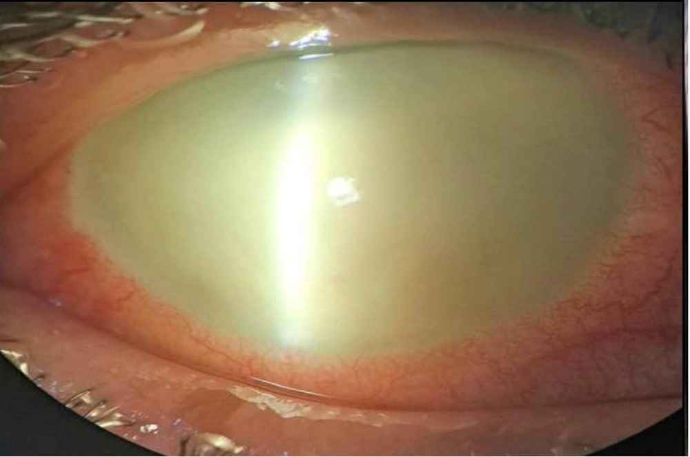
Bacillus cereus is the organism retina surgeons fear most after rural or metallic foreign body injuries, because its toxins can destroy the retina within a day or two. Clues include severe pain, rapid visual loss, a corneal ring infiltrate, and sometimes fever.
Because the course is so fast, treatment should start before cultures return. Vancomycin covers it reliably, so include it in every empirical regimen.
Clinical Signs After Repair
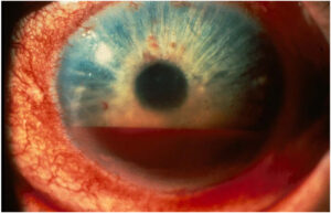
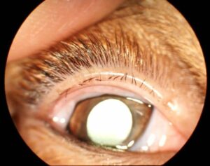
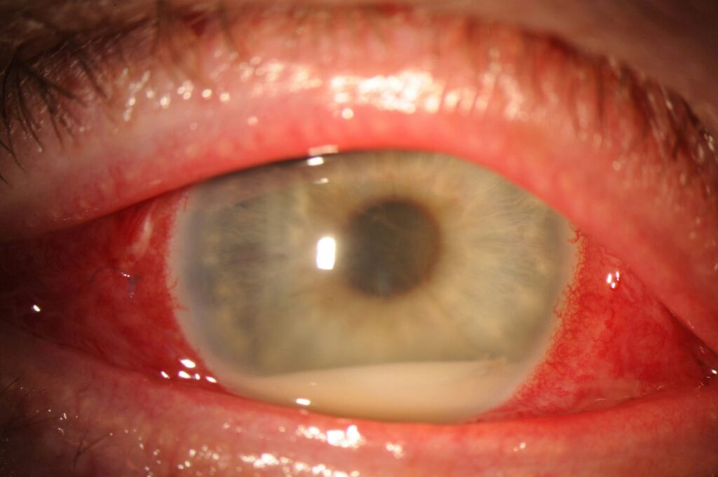
Pain that worsens after the first postoperative day is a warning sign, so next look for hypopyon, anterior chamber fibrin and loss of the red reflex. Lid edema and chemosis that increase out of proportion to the injury also raise concern.
Hyphema, vitreous hemorrhage and traumatic cataract can hide these signs, while sterile uveitis mimics them. Therefore, trend the findings over hours and keep a low threshold for a vitreous tap.
Imaging and Tests in Traumatic Endophthalmitis
B-scan ultrasound shows vitreous opacities and membranes when the media are opaque, although you should avoid pressure on an unrepaired globe. CT localizes metallic foreign bodies and must come before any MRI. Then, vitreous Gram stain and culture confirm the diagnosis and guide therapy.
Prophylactic Antibiotics in Traumatic Endophthalmitis
Many centers give a short course of systemic antibiotics after open globe repair. Oral moxifloxacin reaches useful vitreous levels and covers many trauma organisms, while some units use intravenous vancomycin with ceftazidime, and you should confirm tetanus status in every patient.
Intravitreal Prophylaxis at Repair
Soheilian and colleagues randomized injured eyes to intravitreal vancomycin and ceftazidime or no injection, and the injected group developed endophthalmitis less often. Therefore, many surgeons inject at primary repair when a foreign body or heavy contamination is present. However, the injection carries a small toxicity risk and does not replace prompt foreign body removal.
Treating Established Traumatic Endophthalmitis
Once you suspect infection, take a vitreous sample and inject antibiotics the same day. Standard intravitreal doses are vancomycin 1 mg/0.1 mL and ceftazidime 2.25 mg/0.1 mL.
Amikacin can replace ceftazidime, although it carries a risk of macular infarction. Then, consider repeat injection if the eye fails to improve within 48 hours.
When to Perform Vitrectomy in Traumatic Endophthalmitis
The Endophthalmitis Vitrectomy Study excluded trauma, so its vision-based rule does not apply here (see Endophthalmitis Vitrectomy Study). Many surgeons therefore favor early pars plana vitrectomy after injury. Surgery removes organisms, toxins and the foreign body in one session, and it also clears the media and allows repair of retinal breaks.
Limits and Risks of Surgery
Infected, necrotic retina tears easily during vitrectomy, so confirm the infusion cannula tip in the vitreous cavity before opening it. Corneal haze may require a temporary keratoprosthesis or endoscopic viewing. Even so, surgery cannot restore retina that toxins have already destroyed.
Outcomes and Counseling in Traumatic Endophthalmitis
Visual results are generally worse than after postoperative endophthalmitis. Bacillus infection often ends with no light perception or loss of the eye. As a result, explain early that saving the globe may be the first realistic goal, and discuss sympathetic ophthalmia before deciding to keep a blind, damaged eye.
Pitfalls to Avoid
- Do not blame rising pain after repair on the trauma alone
- Never delay CT when a foreign body is possible
- Avoid MRI until metal is excluded
- Do not apply the EVS vision rule to injured eyes
- Do not stop prophylaxis early after a rural or foreign body injury


Document what you see
Two smartphone imaging tools built for everyday clinic use — one for the slit lamp, one for the fundus.
From Choroida — the team behind this siteReferences
- Essex RW, Yi Q, Charles PG, Allen PJ. Post-traumatic endophthalmitis. Ophthalmology. 2004;111:2015-2022.
- Thompson JT, Parver LM, Enger CL, et al. Infectious endophthalmitis after penetrating injuries with retained intraocular foreign bodies. Ophthalmology. 1993;100:1468-1474.
- Soheilian M, Rafati N, Mohebbi MR, et al. Prophylaxis of acute posttraumatic bacterial endophthalmitis: a multicenter, randomized clinical trial of intraocular antibiotic injection, report 2. Arch Ophthalmol. 2007;125:460-465.
- Endophthalmitis Vitrectomy Study Group. Results of the Endophthalmitis Vitrectomy Study. Arch Ophthalmol. 1995;113:1479-1496.
- Ahmed Y, Schimel AM, Pathengay A, et al. Endophthalmitis following open-globe injuries. Eye (Lond). 2012;26:212-217.

Writes and reviews the clinical articles on Educate. Every post is checked against peer-reviewed sources before it goes live.